
988368
Wed, Oct 21, 2026

Volume 24, Issue 2 (June 2026)
Iranian Rehabilitation Journal 2026, 24(2): 193-206 |
Back to browse issues page




Download citation:
BibTeX | RIS | EndNote | Medlars | ProCite | Reference Manager | RefWorks
Send citation to:


BibTeX | RIS | EndNote | Medlars | ProCite | Reference Manager | RefWorks
Send citation to:
Rezaei M, Soleimanzadeh E, Adigozali H, Ahmadi Kahjoogh M, Jahanjoo F. Contralateral Effects of Dry Needling on Upprt-trapezius Muscle Characteristics and Pain in Patients With Myofascial Pain Syndrome: A Pre-post Quasi-experimental Study. Iranian Rehabilitation Journal 2026; 24 (2) :193-206
URL: http://irj.uswr.ac.ir/article-1-2646-en.html
URL: http://irj.uswr.ac.ir/article-1-2646-en.html
Mandana Rezaei1  , Elham Soleimanzadeh1 , Hakimeh Adigozali *1 , Mina Ahmadi Kahjoogh2 , Fatemeh Jahanjoo3
, Elham Soleimanzadeh1 , Hakimeh Adigozali *1 , Mina Ahmadi Kahjoogh2 , Fatemeh Jahanjoo3
1- Department of Physiotherapy, Faculty of Rehabilitation Sciences, Tabriz University of Medical Sciences, Tabriz, Iran.
2- Department of Occupational Therapy, Faculty of Rehabilitation Sciences, Tabriz University of Medical Sciences, Tabriz, Iran.
3- Physical Medicine and Rehabilitation Research Center, Tabriz Univery of Medical Sciences, Tabriz, Iran.
2- Department of Occupational Therapy, Faculty of Rehabilitation Sciences, Tabriz University of Medical Sciences, Tabriz, Iran.
3- Physical Medicine and Rehabilitation Research Center, Tabriz Univery of Medical Sciences, Tabriz, Iran.
Keywords: Myofascial trigger point, Dry needling, Muscle stiffness, Pain, Ultrasonography, Regional blood flow
Full-Text [PDF 1956 kb]
(288 Downloads)
| Abstract (HTML) (1772 Views)
 .
.
Full-Text: (167 Views)
Introduction
Myofascial pain syndrome (MPS) represents a prevalent and clinically relevant contributor to musculoskeletal pain, with clinical studies reporting high frequencies of MPS and associated myofascial trigger points (MTrPs) among individuals presenting with neck pain [1]. MTrPs are characterized as tender nodules within a taut band of skeletal muscle fibres and commonly reproduce referral pain [1]. The MTrPs exhibit distinct neurobiological characteristics and can lead to abnormalities in the nervous system [2, 3]. The presence of MTrPs often results in increased muscle stiffness, limited range of motion (ROM), and multiple autonomic disturbances [4]. The upper trapezius (UT) muscle, which shows the greatest frequency of MTrPs, is frequently associated with neck pain and impaired upper limb function [5].
MTrPs manifest as palpable nodules within a taut band of skeletal muscle fibres, typically eliciting referred pain upon palpation [1]. The point structures exhibit distinct neurophysiological properties that can disrupt both peripheral and central nervous system functions [2, 3]. MTrPs can contribute to increased muscle stiffness, restricted ROM, and various autonomic dysfunctions [4], with the UT muscle bearing the most significant MTrP prevalence—frequently implicated in neck pain and upper extremity functional disability [5].
In MPS, an abnormal increase in acetylcholine release at the motor end plate within a muscle results in consistent, somatic contraction, which may explain the formation of a taut band [5]. In addition, the higher prevalence of end-plate noise, named ‘spontaneous electrical activity’, is correlated with MTrP excitability [6]. This phenomenon can further lead to an “energy crisis” characterized by increased metabolism and local hypoxia and ischemia within the muscle [7]. Following ischemia, the secretion of vasoactive and algogenic substances alters the threshold of sensory neurons, sensitizing peripheral pain receptors and ultimately affecting central sensitivity [2]. Additionally, hypersensitivity and consistent contraction in the affected muscle alter the viscoelastic properties of the tissue, making active MTrPs stiffer than normal tissues [8]. Therefore, the features of fascia and muscle thickness differ in individuals with MPS compared to healthy subjects [9].
Dry needling effectively deactivates MTrPs immediately after a single session [10]. A meta-analysis further showed its short-term superiority over sham or placebo treatments in reducing neck pain and related disability [11]. When performed properly, it triggers a local twitch response (LTR), recognized as an automatic neuromuscular reaction mediated through spinal mechanisms [12]. This response involves a brief reflex contraction of muscle fibers within the MTrP and is considered a key indicator of effective needle placement. The elicitation of LTR has been suggested to contribute to both localized and generalized therapeutic effects of dry needling [13].
The exact therapeutic effects of dry needling and the underlying mechanisms are not yet completely clarified, but according to some reports, dry needling may lead to suppression of spontaneous electrical activity at the motor end plate [6], reduced muscle stiffness [14], and improved muscle blood flow and oxygenation [8, 15]. These physiological responses are thought to be, at least in part, associated with the reflex muscle contraction induced by the LTR, which may facilitate normalization of neuromuscular and microvascular function. In addition, central sensitization may be one possible explanation for MTrP activation in response to stimulation. A link exists between spinal cord sensory neurons and MTrPs, known as an “MTrP circuit.” Following a strong stimulus from MTrP, the circuit corresponding to this MTrP may enhance the activation of the spinal cord. This phenomenon may be responsible for central sensitization and explain the effects of dry needling. Finally, it is also hypothesized that this method has analgesic effects [2].
Beyond the localized impact of dry needling on MTrPs, limited research has explored its distant effects [15, 16]. Evidence indicates that dry needling can decrease excitability in MTrPs located in remote muscles—either proximal or distal—on the same side of the body [17, 18]. Findings regarding contralateral effects remain inconsistent: certain investigations have documented benefits on the opposite side [6, 16], while others found no effects extending beyond the treated area [18]. Animal research involving dry needling of the gastrocnemius muscle demonstrated reduced endplate noise amplitude in the contralateral gastrocnemius [6].
In contrast, another experimental study in rabbits found no changes in the endplate noise amplitude in the contralateral biceps femoris muscle [18]. The contralateral effects of dry needling applied to infraspinatus muscle MTrPs in individuals with bilateral shoulder pain were examined in another study. This study demonstrated no significant changes in the measured parameters, including shoulder ROM and pain sensitivity in the opposite-side muscle [19].
Dry needling is also effective in improving blood flow and oxygenation in the treated area [8, 20]. In patients with temporomandibular disorder, dry needling of masseter MTrPs elevated oxygen levels in the masseter muscles compared to sham treatment [21].
Nevertheless, evidence on alterations in muscle blood flow at distant sites following dry needling remains inconsistent. Some studies have documented remote circulatory improvements in untreated regions after the intervention [15, 22]. In contrast, additional studies observed no alterations in blood flow within regions remote from the needling site [16, 23]. A significant enhancement in blood flow to the contralateral UT muscle was also observed after dry needling in healthy participants [24]. In addition, the results of another study on patients with fibromyalgia showed similar findings in this regard [15]. However, a reduction in blood flow and oxygen saturation had previously been demonstrated in the contralateral UT muscle of healthy participants after applying dry needling, 5 and 15 minutes later [16].
In clinical practice, most patients with MPS complain of pain in the UT muscle. Furthermore, it may sometimes be impossible to perform direct intervention on the painful area due to pathologic lesions, skin issues, or other relevant problems [25]. Therefore, using techniques at a distant site away from the primary muscle involved in MTrP would be beneficial and may provide an alternative, valuable treatment for symptom relief. To develop better remote objective outcomes with dry needling, it is necessary to simultaneously measure clinical and ultrasonographic variables. Ultrasonographic measurements provide reproducible and reliable dynamic evaluations of muscle morphology and circulatory changes [26]. Within the scope of available evidence, the existing literature provides limited evidence regarding the contralateral effects of dry needling on muscle mechanical properties, such as muscle stiffness. On muscle morphology, including muscle thickness and intramuscular blood circulation, and no consensus has been reached. Based on the proposed neurophysiological mechanisms, including the elicitation of an LTR and central modulation of neuromuscular function, it is plausible to hypothesize that dry needling of the UT may induce measurable contralateral effects. Therefore, this study was designed to simultaneously evaluate clinical and ultrasonographic outcomes, including pain irritability, muscle thickness, stiffness, and blood circulation in the contralateral UT of patients with MPS.
Materials and Methods
Research design and participants
The current study was a prepost quasi-experimental research study. Participants with pain in both shoulders were invited through announcements at the Tabriz University of Medical Sciences’ rehabilitation clinics from September 2021 to February 2022.
Sample size estimation was conducted a priori using G*Power software, version 3.1.9.2 (Universität Kiel, Germany) for a paired t test, based on a moderate effect size of 0.7, α=0.05, and 80% power. Previous findings informed the effect size on pain as the primary outcome [27]. According to this calculation, 14 participants were required.
Thus, 14 patients diagnosed with MPS participated in this study. Participants met the inclusion criteria of right-hand dominance, bilateral shoulder pain, active MTrPs in the UT muscles, age between 20 and 45 years, and normal body mass index (BMI) (18.5–24.9). The exclusion criteria were fibromyalgia syndrome, prior MTrP injections in the UT muscle, needle phobia, history of cancer, upper limb surgery or fracture, radiculopathy, pregnancy, or anticoagulant use [28, 29].
Procedures and interventions
The eligibility of 22 subjects with MPS was assessed prior to inclusion in this study. Inclusion required active MTrPs in the UT muscles, leading to the final enrollment of 14 participants. The study was completed by all 14 participants who were initially enrolled, and no dropouts or missing data occurred; therefore, analyses were conducted on complete cases. First, the MTrP diagnosis was performed. Then, a blinded physiotherapist conducted all evaluations of the study outcomes for both right and left sides. Subsequently, another physiotherapist performed the intervention. All participants received their intervention in the right UT muscle in a single session. After the treatment session, the same assessor recorded the outcome measures on both sides.
MTrP diagnosis
Active MTrPs in both UT muscles were confirmed using the International Consensus criteria [30]. Identification involved finding a palpable taut band with a hypersensitive knot, elicitation of typical pain on compression, and occasionally a LTR. MTrPs were considered active if palpation provoked the patient’s characteristic symptoms, whether partial or complete, including spontaneous or movement-induced pain [30]. The evaluations were conducted by an expert physical therapist in MPS and dry needling, with an average of 14 years of clinical experience in these fields. Once the exact locations of MTrPs had been identified, the skin was marked with permanent ink for assessment.
Outcome measures
After the initial diagnosis of MTrPs, clinical evaluations were performed on both sides, measuring outcomes such as PPT and UT muscle pain intensity. Also, ultrasonographic imaging was performed on both sides to examine the UT muscle thickness, muscle stiffness, and muscle blood flow via grey-scale, color doppler imaging, and ultrasound strain elastography, respectively.
Pain intensity
Once the precise locations of active MTrPs in the bilateral UT muscles were identified, pain intensity was assessed using the visual analog scale (VAS). Participants marked their pain levels on this tool, which consists of a 0-10 range (0=no pain and 10=the worst possible pain). The evaluation was performed pre- and post-intervention, with the mean of three readings from each side used in the statistical analysis [31].
The pain pressure threshold
PPT at the MTrPs in the UT muscles was measured using a digital algometer (J-Tec medical commander power track, US) prior to and following the treatment session. A review has confirmed the high reliability of PPT assessments in the UT with digital algometry [32]. As a measurement tool, the J-tech algometer is a valid instrument for assessing PPT across different regions in a test-retest reliability design [33]. The algometer was placed on the predetermined points of the UT muscle, and pressure was slowly applied. When participants felt pain, they were instructed to respond with “yes”. The amount of pressure was recorded whenever a pain sensation replaced the pressure sensation. This test was performed on both sides three times, and the average of these attempts at each side before and after the dry needling intervention was recorded as PPT. A 30-second pause was implemented between the examinations. Participants were first familiarized with the algometer procedure by applying it to an unaffected muscle [31].
Ultrasonographic assessment
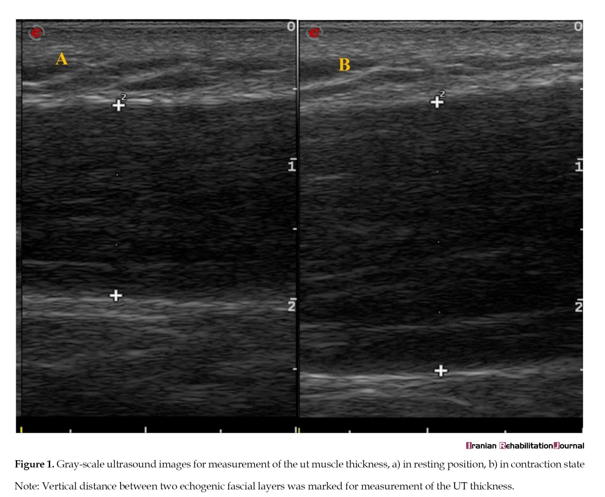
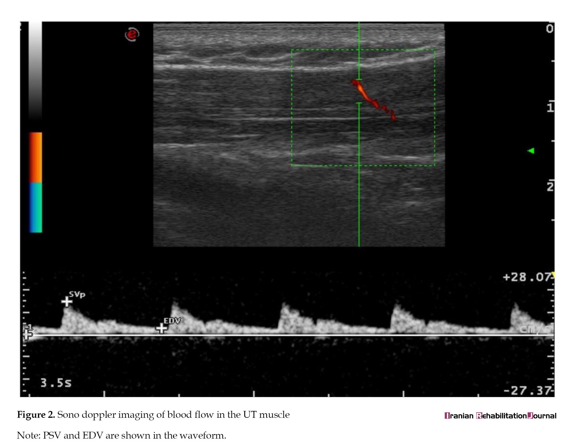
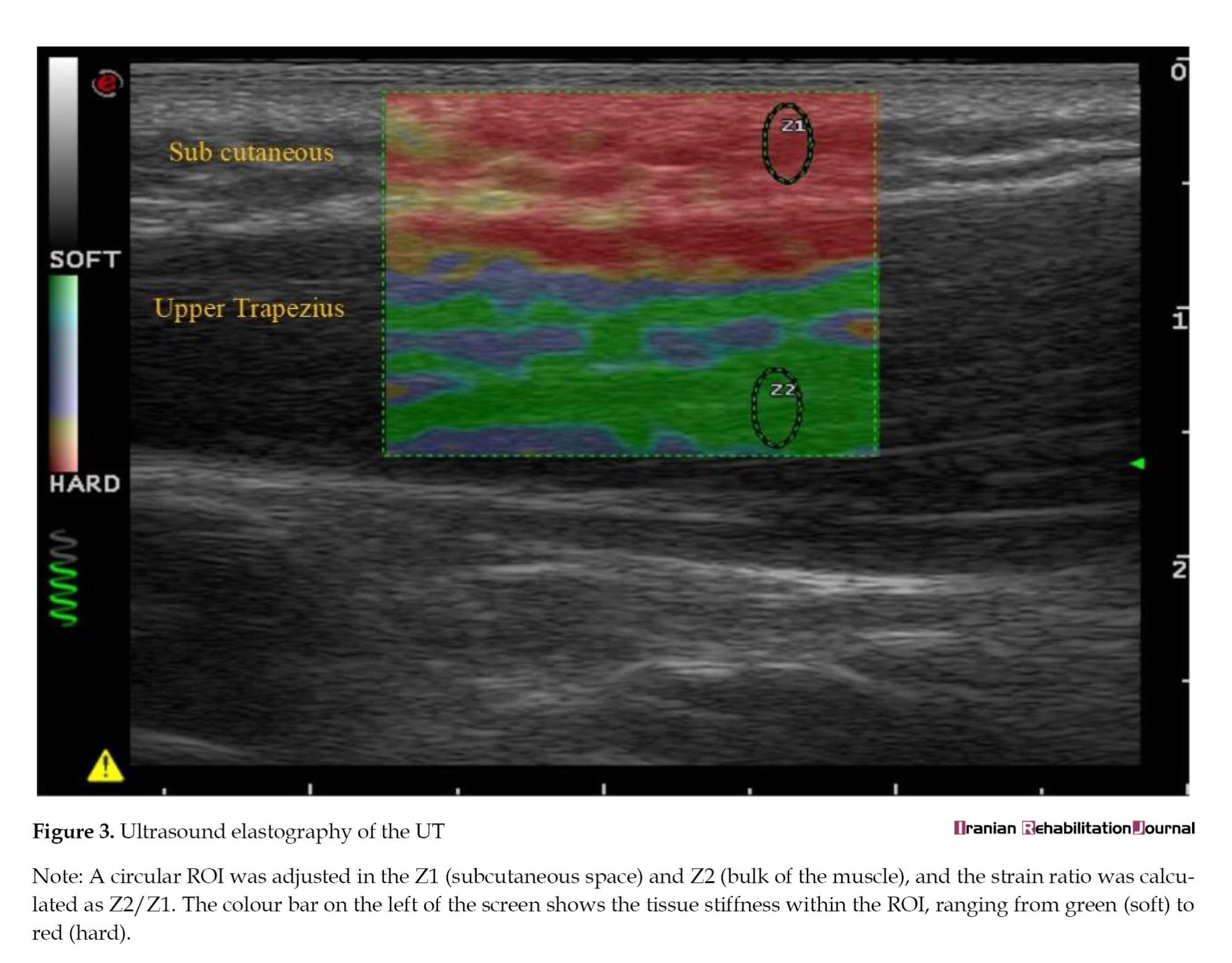
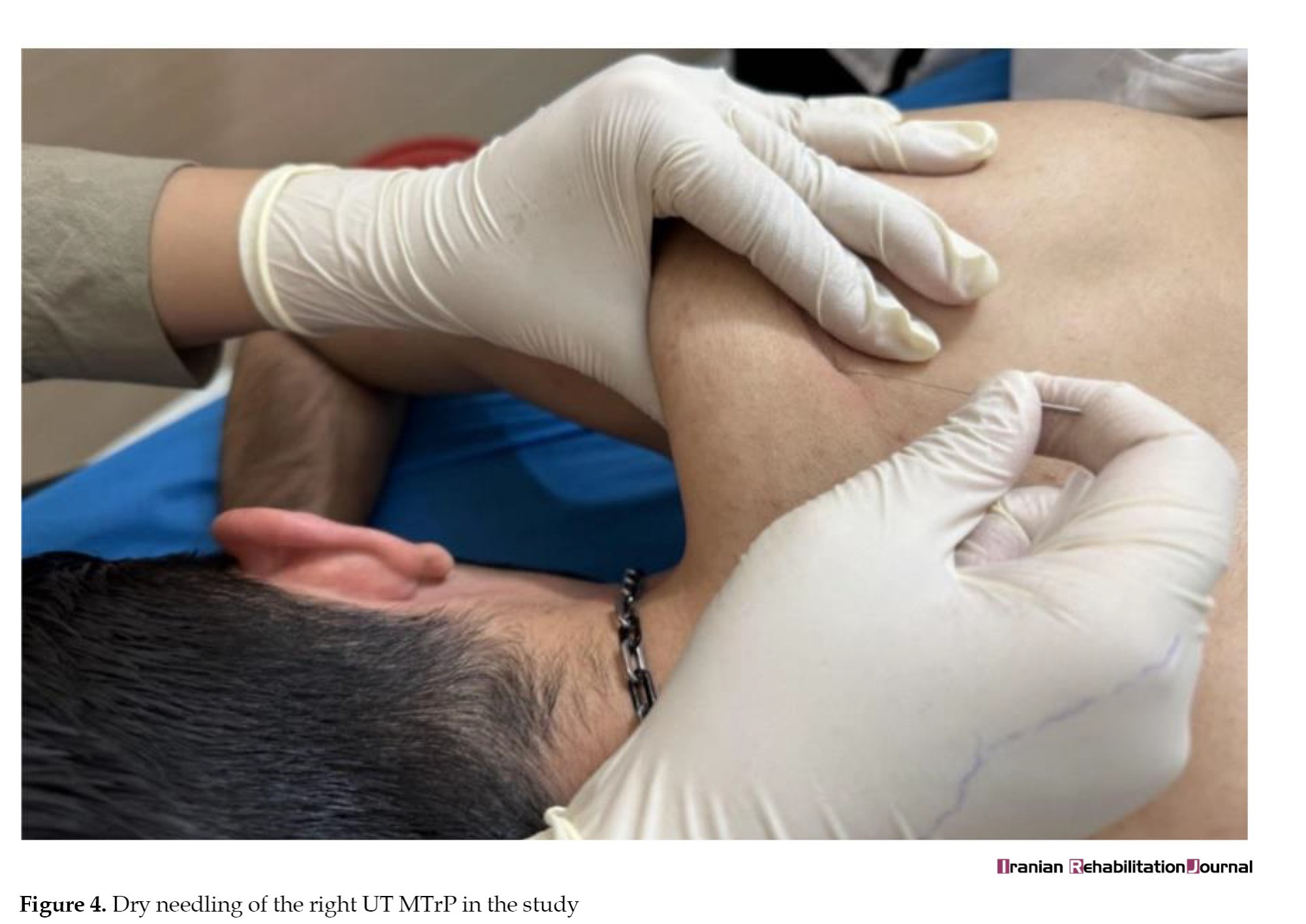
Prior to and following the dry needling intervention, a radiologist assessed the UT muscle on both sides using B-mode imaging, sonoelastography, and Doppler measurements (Figures 1, 2, 3 and 4).
Myofascial pain syndrome (MPS) represents a prevalent and clinically relevant contributor to musculoskeletal pain, with clinical studies reporting high frequencies of MPS and associated myofascial trigger points (MTrPs) among individuals presenting with neck pain [1]. MTrPs are characterized as tender nodules within a taut band of skeletal muscle fibres and commonly reproduce referral pain [1]. The MTrPs exhibit distinct neurobiological characteristics and can lead to abnormalities in the nervous system [2, 3]. The presence of MTrPs often results in increased muscle stiffness, limited range of motion (ROM), and multiple autonomic disturbances [4]. The upper trapezius (UT) muscle, which shows the greatest frequency of MTrPs, is frequently associated with neck pain and impaired upper limb function [5].
MTrPs manifest as palpable nodules within a taut band of skeletal muscle fibres, typically eliciting referred pain upon palpation [1]. The point structures exhibit distinct neurophysiological properties that can disrupt both peripheral and central nervous system functions [2, 3]. MTrPs can contribute to increased muscle stiffness, restricted ROM, and various autonomic dysfunctions [4], with the UT muscle bearing the most significant MTrP prevalence—frequently implicated in neck pain and upper extremity functional disability [5].
In MPS, an abnormal increase in acetylcholine release at the motor end plate within a muscle results in consistent, somatic contraction, which may explain the formation of a taut band [5]. In addition, the higher prevalence of end-plate noise, named ‘spontaneous electrical activity’, is correlated with MTrP excitability [6]. This phenomenon can further lead to an “energy crisis” characterized by increased metabolism and local hypoxia and ischemia within the muscle [7]. Following ischemia, the secretion of vasoactive and algogenic substances alters the threshold of sensory neurons, sensitizing peripheral pain receptors and ultimately affecting central sensitivity [2]. Additionally, hypersensitivity and consistent contraction in the affected muscle alter the viscoelastic properties of the tissue, making active MTrPs stiffer than normal tissues [8]. Therefore, the features of fascia and muscle thickness differ in individuals with MPS compared to healthy subjects [9].
Dry needling effectively deactivates MTrPs immediately after a single session [10]. A meta-analysis further showed its short-term superiority over sham or placebo treatments in reducing neck pain and related disability [11]. When performed properly, it triggers a local twitch response (LTR), recognized as an automatic neuromuscular reaction mediated through spinal mechanisms [12]. This response involves a brief reflex contraction of muscle fibers within the MTrP and is considered a key indicator of effective needle placement. The elicitation of LTR has been suggested to contribute to both localized and generalized therapeutic effects of dry needling [13].
The exact therapeutic effects of dry needling and the underlying mechanisms are not yet completely clarified, but according to some reports, dry needling may lead to suppression of spontaneous electrical activity at the motor end plate [6], reduced muscle stiffness [14], and improved muscle blood flow and oxygenation [8, 15]. These physiological responses are thought to be, at least in part, associated with the reflex muscle contraction induced by the LTR, which may facilitate normalization of neuromuscular and microvascular function. In addition, central sensitization may be one possible explanation for MTrP activation in response to stimulation. A link exists between spinal cord sensory neurons and MTrPs, known as an “MTrP circuit.” Following a strong stimulus from MTrP, the circuit corresponding to this MTrP may enhance the activation of the spinal cord. This phenomenon may be responsible for central sensitization and explain the effects of dry needling. Finally, it is also hypothesized that this method has analgesic effects [2].
Beyond the localized impact of dry needling on MTrPs, limited research has explored its distant effects [15, 16]. Evidence indicates that dry needling can decrease excitability in MTrPs located in remote muscles—either proximal or distal—on the same side of the body [17, 18]. Findings regarding contralateral effects remain inconsistent: certain investigations have documented benefits on the opposite side [6, 16], while others found no effects extending beyond the treated area [18]. Animal research involving dry needling of the gastrocnemius muscle demonstrated reduced endplate noise amplitude in the contralateral gastrocnemius [6].
In contrast, another experimental study in rabbits found no changes in the endplate noise amplitude in the contralateral biceps femoris muscle [18]. The contralateral effects of dry needling applied to infraspinatus muscle MTrPs in individuals with bilateral shoulder pain were examined in another study. This study demonstrated no significant changes in the measured parameters, including shoulder ROM and pain sensitivity in the opposite-side muscle [19].
Dry needling is also effective in improving blood flow and oxygenation in the treated area [8, 20]. In patients with temporomandibular disorder, dry needling of masseter MTrPs elevated oxygen levels in the masseter muscles compared to sham treatment [21].
Nevertheless, evidence on alterations in muscle blood flow at distant sites following dry needling remains inconsistent. Some studies have documented remote circulatory improvements in untreated regions after the intervention [15, 22]. In contrast, additional studies observed no alterations in blood flow within regions remote from the needling site [16, 23]. A significant enhancement in blood flow to the contralateral UT muscle was also observed after dry needling in healthy participants [24]. In addition, the results of another study on patients with fibromyalgia showed similar findings in this regard [15]. However, a reduction in blood flow and oxygen saturation had previously been demonstrated in the contralateral UT muscle of healthy participants after applying dry needling, 5 and 15 minutes later [16].
In clinical practice, most patients with MPS complain of pain in the UT muscle. Furthermore, it may sometimes be impossible to perform direct intervention on the painful area due to pathologic lesions, skin issues, or other relevant problems [25]. Therefore, using techniques at a distant site away from the primary muscle involved in MTrP would be beneficial and may provide an alternative, valuable treatment for symptom relief. To develop better remote objective outcomes with dry needling, it is necessary to simultaneously measure clinical and ultrasonographic variables. Ultrasonographic measurements provide reproducible and reliable dynamic evaluations of muscle morphology and circulatory changes [26]. Within the scope of available evidence, the existing literature provides limited evidence regarding the contralateral effects of dry needling on muscle mechanical properties, such as muscle stiffness. On muscle morphology, including muscle thickness and intramuscular blood circulation, and no consensus has been reached. Based on the proposed neurophysiological mechanisms, including the elicitation of an LTR and central modulation of neuromuscular function, it is plausible to hypothesize that dry needling of the UT may induce measurable contralateral effects. Therefore, this study was designed to simultaneously evaluate clinical and ultrasonographic outcomes, including pain irritability, muscle thickness, stiffness, and blood circulation in the contralateral UT of patients with MPS.
Materials and Methods
Research design and participants
The current study was a prepost quasi-experimental research study. Participants with pain in both shoulders were invited through announcements at the Tabriz University of Medical Sciences’ rehabilitation clinics from September 2021 to February 2022.
Sample size estimation was conducted a priori using G*Power software, version 3.1.9.2 (Universität Kiel, Germany) for a paired t test, based on a moderate effect size of 0.7, α=0.05, and 80% power. Previous findings informed the effect size on pain as the primary outcome [27]. According to this calculation, 14 participants were required.
Thus, 14 patients diagnosed with MPS participated in this study. Participants met the inclusion criteria of right-hand dominance, bilateral shoulder pain, active MTrPs in the UT muscles, age between 20 and 45 years, and normal body mass index (BMI) (18.5–24.9). The exclusion criteria were fibromyalgia syndrome, prior MTrP injections in the UT muscle, needle phobia, history of cancer, upper limb surgery or fracture, radiculopathy, pregnancy, or anticoagulant use [28, 29].
Procedures and interventions
The eligibility of 22 subjects with MPS was assessed prior to inclusion in this study. Inclusion required active MTrPs in the UT muscles, leading to the final enrollment of 14 participants. The study was completed by all 14 participants who were initially enrolled, and no dropouts or missing data occurred; therefore, analyses were conducted on complete cases. First, the MTrP diagnosis was performed. Then, a blinded physiotherapist conducted all evaluations of the study outcomes for both right and left sides. Subsequently, another physiotherapist performed the intervention. All participants received their intervention in the right UT muscle in a single session. After the treatment session, the same assessor recorded the outcome measures on both sides.
MTrP diagnosis
Active MTrPs in both UT muscles were confirmed using the International Consensus criteria [30]. Identification involved finding a palpable taut band with a hypersensitive knot, elicitation of typical pain on compression, and occasionally a LTR. MTrPs were considered active if palpation provoked the patient’s characteristic symptoms, whether partial or complete, including spontaneous or movement-induced pain [30]. The evaluations were conducted by an expert physical therapist in MPS and dry needling, with an average of 14 years of clinical experience in these fields. Once the exact locations of MTrPs had been identified, the skin was marked with permanent ink for assessment.
Outcome measures
After the initial diagnosis of MTrPs, clinical evaluations were performed on both sides, measuring outcomes such as PPT and UT muscle pain intensity. Also, ultrasonographic imaging was performed on both sides to examine the UT muscle thickness, muscle stiffness, and muscle blood flow via grey-scale, color doppler imaging, and ultrasound strain elastography, respectively.
Pain intensity
Once the precise locations of active MTrPs in the bilateral UT muscles were identified, pain intensity was assessed using the visual analog scale (VAS). Participants marked their pain levels on this tool, which consists of a 0-10 range (0=no pain and 10=the worst possible pain). The evaluation was performed pre- and post-intervention, with the mean of three readings from each side used in the statistical analysis [31].
The pain pressure threshold
PPT at the MTrPs in the UT muscles was measured using a digital algometer (J-Tec medical commander power track, US) prior to and following the treatment session. A review has confirmed the high reliability of PPT assessments in the UT with digital algometry [32]. As a measurement tool, the J-tech algometer is a valid instrument for assessing PPT across different regions in a test-retest reliability design [33]. The algometer was placed on the predetermined points of the UT muscle, and pressure was slowly applied. When participants felt pain, they were instructed to respond with “yes”. The amount of pressure was recorded whenever a pain sensation replaced the pressure sensation. This test was performed on both sides three times, and the average of these attempts at each side before and after the dry needling intervention was recorded as PPT. A 30-second pause was implemented between the examinations. Participants were first familiarized with the algometer procedure by applying it to an unaffected muscle [31].
Ultrasonographic assessment
Prior to and following the dry needling intervention, a radiologist assessed the UT muscle on both sides using B-mode imaging, sonoelastography, and Doppler measurements (Figures 1, 2, 3 and 4).
.Based on our previous studies, good to excellent reliability (intraclass correlation coefficient >0.806) was reported for ultrasonographic scanning of the UT muscle, including muscle thickness, intramuscular blood flow, and muscle stiffness, in patients with MPS and healthy participants [26, 34].
Muscle thickness in the UT of each participant was assessed using a Siemens Acuson Juniper diagnostic ultrasound system (Siemens Healthcare, Erlangen, Germany) equipped with a 3-12 MHz linear probe. Probe pressure was standardized by applying the minimum pressure required to obtain clear, stable images while avoiding visible tissue compression. Pressure consistency was visually verified using the pressure indicator shown alongside the representative ultrasound images (Figure 3). For ultrasonographic scanning, the participants assumed an upright seated position in a chair, and the scanning location on the muscle was a previously marked site. Using B-mode grey-scale imaging, muscle thickness was determined as the vertical distance between the echogenic fascial layers of the UT, with the ultrasound probe positioned along the fibers of the UT. The thickness was assessed in a resting state and during contraction. In the resting state, participants assumed a comfortable seated posture, with their shoulders abducted to 0°, and their forearms resting on the chair armrests [8]. During muscle contraction, participants were asked to perform 90° shoulder abduction combined with elbow extension and pronation, holding this position for 10 seconds (Figure 1).
Following this, color Doppler ultrasonography was performed on each participant’s UT muscle at the pre-marked location to assess intramuscular blood flow. Spectral Doppler waveforms, which indicate blood flow velocity, were analyzed to calculate key parameters per cardiac cycle: Peak systolic velocity (PSV), end-diastolic velocity (EDV), and resistance index (RI) [8]. In each waveform, the left limit reflects the onset of systole, the high point of the wave corresponds with peak systole, and the right limit demonstrates the end of diastole. The RI, a standard measure in vascular assessment, was calculated as the ratio of the difference between PSV and EDV to PSV [35]. In the study, muscle blood flow parameters were measured in the arterioles or arteries located near the MTrP of the UT muscles on both sides (Figure 2).
Finally, muscle stiffness was measured using an ultrasound elastogram. Tissue deformation is shown as a color-coded map on a grey-scale scale, known as an elastogram. Strain-ratio measurements provide semi-quantitative data on tissue deformation within an elastogram. This ratio represents the difference in deformation between the two user-defined areas under a compressive force. Soft areas exhibit higher strain values than stiff areas. To obtain the UT muscle strain ratio, participants were asked to sit upright in a comfortable position. The ultrasound probe was oriented perpendicular to the previously marked site of the UT muscle and then compressed rhythmically with rhythmic, vertical cyclic compressions (with a frequency of 2 Hz). The pressure indicator item in the ultrasonogram controlled the manual compression force. Subsequently, strain elastography was applied by superimposing a color-coded elasticity map onto the B-mode ultrasound images. This mapping was generated based on the degree of tissue deformation (strain) induced during probe compression, where softer tissues appeared in green and progressively stiffer tissues transitioned to red. Once the most representative image was selected, two distinct regions of interest (ROIs) were precisely delineated to enable strain-ratio quantification. The first ROI served as the reference point, as positioned within the adjacent subcutaneous tissue. The second ROI was carefully placed in the immediate vicinity of the identified MTrP within the UT muscle. To ensure measurement reliability and comparability, both ROIs were maintained at identical dimensions for all participants, regardless of whether they represented the reference point or the target lesion site [8]. The strain ratio was calculated between two selected areas, and a decreased strain ratio implied increased muscle stiffness [36] (Figure 3).
Interventions
In this study, dry needling was applied to the MTrP in the right UT muscle. Subjects were positioned in the prone position. Prior to the procedure, the skin over the targeted area in the right UT muscle was cleaned using an alcohol pad. The therapist held the UT muscle to locate the needle. A sterilized stainless-steel needle, 50 mm in length and 0.25 mm in diameter (Dong–Bang, Korea), was deeply applied into the MTrP to produce LTR. Correct needle placement was confirmed by eliciting the familiar referred pain pattern along with an LTR. The needle was then rapidly manipulated using a fast-in, fast-out technique. Manipulation continued until no further LTRs could be provoked, at which point the needle was removed [29]. The intervention lasted 2 minutes (Figure 4).
Statistical analysis
Frequencies and percentages were used to describe qualitative data, while Mean±SD summarized quantitative data. The normality of all variables was confirmed using the Shapiro-Wilk test. All outcomes were analyzed (within group) using paired t tests for the treatment and untreated sides separately. Effect sizes were calculated using Cohen’s d for paired comparisons to quantify the magnitude of change, with conventional benchmarks (e.g. d≈0.2=small, 0.5=medium, 0.8=large). All analyses were performed using SPSS software, version 22.0, with P<0.05 considered statistically significant.
Results
Table 1 presents the descriptive statistics for all participants.
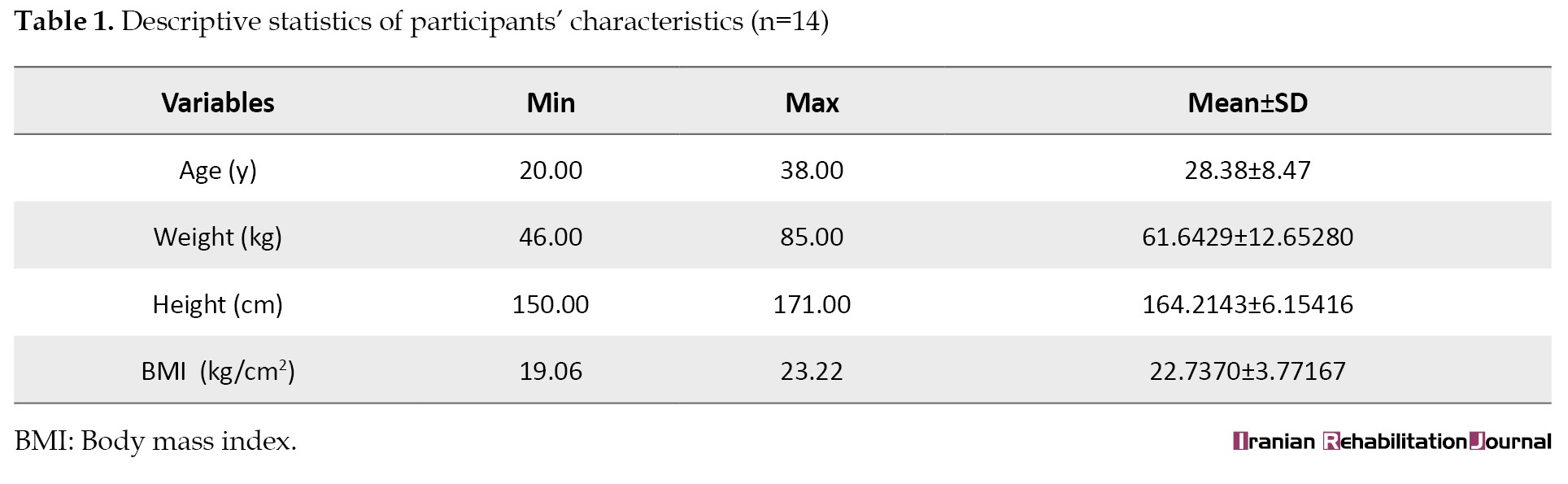
The participants ranged in age from 20 to 38 years, with a mean age of 28.38±8.47 years. The BMI values ranged from 19.06 to 23.22, indicating that all participants fell within the normal weight range and were neither overweight nor obese. Given the study’s sample size, we used the Shapiro-Wilk test to assess normality. The results indicated that all measured variables were normally distributed. The initial measurements for both sides were comparable and showed no notable discrepancies, as indicated by a P>0.001. Dry needling of the UT muscle resulted in significant improvements in the VAS score, PPT, muscle thickness at rest, PSV, EDV, and muscle stiffness on the treated side (P<0.001).
Furthermore, the effects of dry needling were significant for VAS, PPT, muscle thickness at rest, and PSV of the contralateral UT muscle (P<0.05). In contrast, changes in EDV, RI, and strain ratio did not reach statistical significance (Table 2).
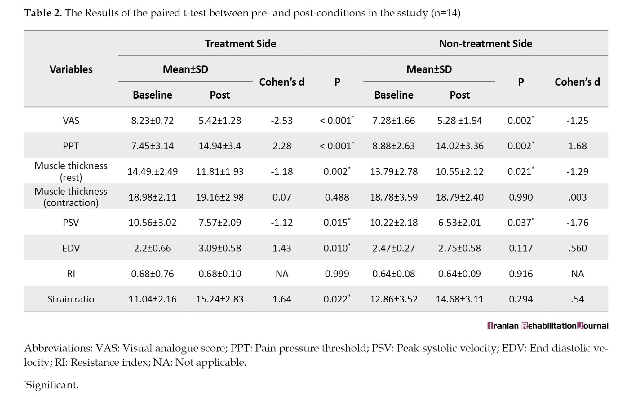
The results of the between-group comparisons revealed that the improvement in VAS scores was significantly greater on the treatment side than on the contralateral side (P<0.001). Similarly, muscle stiffness exhibited a notable improvement on the treatment side (P=0.005).
The reduction in VAS on the treated side corresponded to Cohen’s d of –2.53, indicating a considerable effect size and very substantial clinical improvement in pain intensity. On the untreatment side, Cohen’s d=–1.25 also reflected a substantial effect, albeit smaller than on the treated side. Similarly, PPT demonstrated large effect sizes bilaterally (Table 2).
Discussion
In the present study, significant improvements were observed in both subjective and objective measurements on both treated and untreated sides after right UT dry needling. On the treated ipsilateral side, reductions in pain intensity, resting muscle thickness, and muscle stiffness were observed, along with an increase in pain threshold and muscle blood flow. The short-term effects of dry needling on the contralateral side included improvements in pain intensity and threshold, increased muscle blood flow, and reduced muscle thickness.
Pain intensity
Although some factors, such as muscle overuse, muscle overload, and trauma, are considered etiologic agents of MPS, the pathogenesis of this syndrome is elusive [38]. Abnormal release of noxious substances after muscle overuse leads to either sensitizing or activating nociceptors and may set up a consistent pain state [2]. Therefore, muscle pain is a common complaint among active MTrPs. In the present study, pain reduction was observed on both sides after dry needling. The decrease in pain intensity in the study corresponded to large effect sizes (Cohen’s d=–2.53 for the treated side and –1.25 for the contralateral side), indicating substantial clinical improvement. There are conflicting findings about this issue. Consistent with our study, Gerber et al. (2015) found that dry needling on one side of patients with bilateral MTrPs significantly reduced pain intensity on both the treated and untreated sides [27]. In another study, Ohkubo et al. (2007) showed that dry needling did not considerably affect pain levels of the contralateral infraspinatus muscle’s MTrP [39].
Needling frequency, duration, and needle placement are essential parameters that can alter the effects of this method on clinical symptoms. Mechanical stimulation with dry needling in the MTrP and elicited LTR may explain the beneficial impact of dry needling on pain level [40]. In other words, once LTR altered large-diameter afferent sensory signals projecting to the spinal cord, pain was then blocked via the “gate-controlling” mechanism [3, 5, 41]. Dry needling may activate Aβ, Aδ, and C fibres by releasing biomarkers associated with inflammation. The activated fibres may stimulate supraspinal regions involved in pain modulation, thereby restoring normal central sensitization processes [2].
The pain pressure threshold
In the current study, PPT increased significantly on the ipsilateral side after dry needling. PPT reflects a patient’s perception of pain. Limited evidence suggests that dry needling can increase PPT in treated muscles immediately and at the 12-week post-intervention evaluation [42]. It appears that sensitizing biochemical substances, such as neuropeptides and catecholamines, influences PPT [43]. An LTR with dry needling may reveal alterations in bioactive sensitizing substances [43]. Indeed, increasing local blood flow and subsequently washing out pain mediators, as well as chemical normalization of the MTrP area, may correspond to the observed increase in pain threshold after dry needling [2]. Also, intense stimulation of nociceptors during dry needling of the UT muscle may enhance activity in somatosensory and limbic regions involved in pain processing [3, 44].
Furthermore, the current study demonstrated that PPT significantly increased on the contralateral side after dry needling. The observed increases in PPT corresponded to meaningful effect sizes (Cohen’s d >1.0), suggesting a substantial clinical influence of dry needling on both the ipsilateral and contralateral sides. Previous research has demonstrated that dry needling can reduce pain sensitivity at sites distant from the treated area [31, 45]. Central sensitization may be one possible explanation for MTrP activation in response to remote stimulation. A link exists between spinal cord sensory neurons and MTrPs, known as an “MTrP circuit.” Following a strong stimulus from MTrP, the circuit corresponding to this site may enhance the activation of the other sites in the spinal cord. This phenomenon may be responsible for central sensitization and explain contralateral effects on PPT [45]. Conversely, no significant alterations in contralateral PPT were observed following dry needling in another study [39]. Due to a lack of evidence about this issue, there is a call for further studies.
Ultrasonographic features
To demonstrate the effectiveness of dry needling, objective measures are necessary. Ultrasonography is a practical, non-invasive method for this purpose [22]. It has been reported that the thickness of the muscles affected by MTrP is higher than that of healthy muscles [46]. In this study, dry needling of the UT muscle was associated with a reduction in resting muscle thickness on both sides. The findings of the present investigation are consistent with prior research examining the impact of dry needling on the lumbar multifidus. They observed a reduction in the resting thickness of the transverse abdominis following dry needling of the lumbar multifidus [47]. The impacts of dry needling could be explained by mechanical and neurophysiological mechanisms [48, 49]. In terms of mechanical changes, dry needling may disrupt myofascial knots, induce elongation of contracted cytoskeletal elements while decreasing actin–myosin filament overlap, which may contribute to suppression of excessive muscle activity in the treated region [50, 51].
Furthermore, the effects on the untreated side extend beyond its mechanical effects. From a neurophysiological perspective, a reduction in metabolic mediators and a nociceptive pain barrage, accompanied by a descending pain-inhibitory system, might explain its impact on muscle thickness [49]. Therefore, the reduction in contralateral UT muscle thickness observed in this study may result from pain alleviation and the inhibition of the pain-spasm cycle. In the current study, a decrease in PSV on both sides and an increase in EDV on the treated side after the intervention were observed. Different blood flow waveforms have been reported in muscles with MTrPs [52]. The blood flow pattern near MTrPs shows higher PSV and lower EDV than at the normal site [53]. These changes can be due to muscle contraction, which compresses the arteries and veins near the MTrP. Following these changes, vascular resistance increases, thereby decreasing perfusion to the muscles [16, 53]. Our findings align with those reported in previous studies on various muscles [15, 16, 54]. However, Sandberg et al. showed that blood flow changes on the untreated side were substantially lower than in the stimulated muscle [15]. It should be considered that dry needling is a painful intervention. Therefore, nociceptive afferents are strongly activated during needle manipulation, accompanied by the axonal and vasodilator reflexes, which may enhance blood circulation by releasing vasodilatory biochemical agents (calcitonin gene-related peptide and substance P) [16].
According to Simon’s theory, the sympathetic nervous system is involved in MPS. Changes in the autonomic nervous system, such as the sympathetic stress response, may be another mechanism underlying the increase in blood flow, broadening the stimulated region, possibly mediated by reflex pathways [15]. Alterations in blood flow following dry needling in distant regions have shown inconsistent results across studies [2]. Moreover, some studies have not yielded a significant increase in blood flow in untreated areas after dry needling [16]. The different natures of the diseases and the varying evaluation methods used in these studies may explain the differences in the results.
Musculoskeletal pathologies can affect the viscoelastic properties of muscles [53]. In the study, dry needling led to a substantial decrease in the UT’s stiffness on the treated side. However, on the side opposite to the treatment, the reduction in muscle stiffness was not statistically significant. Earlier studies have demonstrated that dry needling reduces muscle stiffness [8, 14, 55, 56]. There is currently no information regarding the remote effects of this method on the opposite side of treatment. It appears that the decline in muscle stiffness following dry needling is related to the LTR; needle manipulation in MTrP induces a local stretch of the contracted cytoskeletal structures, thereby restoring the resting length [40]. Therefore, such changes require direct needle application.
Conclusion
In conclusion, this study provides preliminary evidence that dry needling, in addition to its direct effects on the treated UT, may induce immediate changes in the contralateral muscle. Observed improvements in pain and sonographic features suggest that dry needling may have effects beyond the treated area. These findings should be interpreted with caution due to the small sample size and single-session intervention. Future studies with larger samples and longer follow-up are warranted to confirm these effects and further explore underlying mechanisms. Dry needling may represent a potential component of treatment protocols for patients with MPS, particularly when direct intervention is not feasible or preferred.
Strengths and limitations
There were some limitations in the study. First, dry needling was applied in a single session, with a pre-post design and no control group. By adding further treatment sessions to a randomized controlled trial, changes in outcome measures can be better understood. Second, the study evaluated the immediate effects of dry needling, and follow-up studies are recommended for future research. Third, we did not design a sham dry-needling group because inserting a needle into the skin can alter analgesic neural responses. Fourth, further studies in this area with larger sample sizes on different muscle groups involved in MPS, as well as in various diseases, such as fibromyalgia, are suggested. Finally, because dominance influences motor control and muscle morphology, we assessed only the right side in right-handed individuals. However, we did not consider the possibility of greater pain on the left side, which may have affected the results. Accordingly, future studies should exclude participants whose non-dominant side is more painful and include only individuals in whom the painful side aligns with the dominant side. This approach may reduce variability related to lateral dominance in motor control and muscle morphology.
Recommendations for future studies
Recent studies have shown growing support for the clinical effectiveness of dry needling in patients with MPS. Nevertheless, although generally supporting the direct effects of dry needling, remote and contralateral effects remain elusive. More trials are necessary to explore the impact of dry needling, particularly in distant areas, using a larger study population. It has been challenging to prove the method’s effectiveness due to the absence of objective metrics. In future studies, we need to combine the remote efficacy of dry needling with objective measures, such as ultrasonography and electromyography.
Overall, from a clinical perspective, the dry needling technique may improve pain, muscle thickness, and blood flow on both the treated and contralateral sides. These results highlight the multifaceted benefits of dry needling for muscle healing and recovery, mediated by distinct mechanisms, in patients with MPS.
Clinical implications
Dry needling targeted at MTrPs in the UT muscle on one side may elicit contralateral therapeutic effects, characterized by reduced pain intensity and alterations in sonographic features on the untreated side, supporting its adjunctive role in the management of MPS. Given these constraints, clinicians should cautiously consider this approach for integration into multimodal protocols addressing bilateral UT involvement, particularly when direct ipsilateral intervention seems impractical (e.g. due to hypersensitivity, anatomical limitations, or patient/clinician preferences).
Ethical Considerations
Compliance with ethical guidelines
This study was approved by the Ethics Committee of Tabriz University of Medical Sciences, Tabriz, Iran (Code: IR.TBZMED.REC.1400.529). The patient was provided with a detailed explanation of the procedure and the purpose of the intervention. Verbal and written consent were obtained before the commencement of the intervention.
Funding
This research didn’t receive any grant from funding agencies in the public, commercial or non-profit sectors.
Authors' contributions
Conceptualization: Hakimeh Adigozali and Elhan Soleimanzadeh; Methodology: Fatemeh Jahanjoo, Mina Ahmadi-Kahjoogh and Mandana Rezaei; Statistical analysis: Mandana Rezaei and Fatemeh Jahanjoo; Interpretation of the results: Mina Ahmadi-Kahjoogh, Hakimeh Adigozali and Elhan Soleimanzadeh; Manuscript writing: Elhan Soleimanzadeh and Hakimeh Adigozali; Manuscript revision: Hakimeh Adigozali.
Conflict of interest
The authors declared no conflicts of interest.
Acknowledgments
The authors thank Clinical Research Development Unit colleagues at Razi Psychiatric Hospital, Tabriz University of Medical Sciences and grateful for their cooperation, collaboration, and support.
Muscle thickness in the UT of each participant was assessed using a Siemens Acuson Juniper diagnostic ultrasound system (Siemens Healthcare, Erlangen, Germany) equipped with a 3-12 MHz linear probe. Probe pressure was standardized by applying the minimum pressure required to obtain clear, stable images while avoiding visible tissue compression. Pressure consistency was visually verified using the pressure indicator shown alongside the representative ultrasound images (Figure 3). For ultrasonographic scanning, the participants assumed an upright seated position in a chair, and the scanning location on the muscle was a previously marked site. Using B-mode grey-scale imaging, muscle thickness was determined as the vertical distance between the echogenic fascial layers of the UT, with the ultrasound probe positioned along the fibers of the UT. The thickness was assessed in a resting state and during contraction. In the resting state, participants assumed a comfortable seated posture, with their shoulders abducted to 0°, and their forearms resting on the chair armrests [8]. During muscle contraction, participants were asked to perform 90° shoulder abduction combined with elbow extension and pronation, holding this position for 10 seconds (Figure 1).
Following this, color Doppler ultrasonography was performed on each participant’s UT muscle at the pre-marked location to assess intramuscular blood flow. Spectral Doppler waveforms, which indicate blood flow velocity, were analyzed to calculate key parameters per cardiac cycle: Peak systolic velocity (PSV), end-diastolic velocity (EDV), and resistance index (RI) [8]. In each waveform, the left limit reflects the onset of systole, the high point of the wave corresponds with peak systole, and the right limit demonstrates the end of diastole. The RI, a standard measure in vascular assessment, was calculated as the ratio of the difference between PSV and EDV to PSV [35]. In the study, muscle blood flow parameters were measured in the arterioles or arteries located near the MTrP of the UT muscles on both sides (Figure 2).
Finally, muscle stiffness was measured using an ultrasound elastogram. Tissue deformation is shown as a color-coded map on a grey-scale scale, known as an elastogram. Strain-ratio measurements provide semi-quantitative data on tissue deformation within an elastogram. This ratio represents the difference in deformation between the two user-defined areas under a compressive force. Soft areas exhibit higher strain values than stiff areas. To obtain the UT muscle strain ratio, participants were asked to sit upright in a comfortable position. The ultrasound probe was oriented perpendicular to the previously marked site of the UT muscle and then compressed rhythmically with rhythmic, vertical cyclic compressions (with a frequency of 2 Hz). The pressure indicator item in the ultrasonogram controlled the manual compression force. Subsequently, strain elastography was applied by superimposing a color-coded elasticity map onto the B-mode ultrasound images. This mapping was generated based on the degree of tissue deformation (strain) induced during probe compression, where softer tissues appeared in green and progressively stiffer tissues transitioned to red. Once the most representative image was selected, two distinct regions of interest (ROIs) were precisely delineated to enable strain-ratio quantification. The first ROI served as the reference point, as positioned within the adjacent subcutaneous tissue. The second ROI was carefully placed in the immediate vicinity of the identified MTrP within the UT muscle. To ensure measurement reliability and comparability, both ROIs were maintained at identical dimensions for all participants, regardless of whether they represented the reference point or the target lesion site [8]. The strain ratio was calculated between two selected areas, and a decreased strain ratio implied increased muscle stiffness [36] (Figure 3).
Interventions
In this study, dry needling was applied to the MTrP in the right UT muscle. Subjects were positioned in the prone position. Prior to the procedure, the skin over the targeted area in the right UT muscle was cleaned using an alcohol pad. The therapist held the UT muscle to locate the needle. A sterilized stainless-steel needle, 50 mm in length and 0.25 mm in diameter (Dong–Bang, Korea), was deeply applied into the MTrP to produce LTR. Correct needle placement was confirmed by eliciting the familiar referred pain pattern along with an LTR. The needle was then rapidly manipulated using a fast-in, fast-out technique. Manipulation continued until no further LTRs could be provoked, at which point the needle was removed [29]. The intervention lasted 2 minutes (Figure 4).
Statistical analysis
Frequencies and percentages were used to describe qualitative data, while Mean±SD summarized quantitative data. The normality of all variables was confirmed using the Shapiro-Wilk test. All outcomes were analyzed (within group) using paired t tests for the treatment and untreated sides separately. Effect sizes were calculated using Cohen’s d for paired comparisons to quantify the magnitude of change, with conventional benchmarks (e.g. d≈0.2=small, 0.5=medium, 0.8=large). All analyses were performed using SPSS software, version 22.0, with P<0.05 considered statistically significant.
Results
Table 1 presents the descriptive statistics for all participants.
The participants ranged in age from 20 to 38 years, with a mean age of 28.38±8.47 years. The BMI values ranged from 19.06 to 23.22, indicating that all participants fell within the normal weight range and were neither overweight nor obese. Given the study’s sample size, we used the Shapiro-Wilk test to assess normality. The results indicated that all measured variables were normally distributed. The initial measurements for both sides were comparable and showed no notable discrepancies, as indicated by a P>0.001. Dry needling of the UT muscle resulted in significant improvements in the VAS score, PPT, muscle thickness at rest, PSV, EDV, and muscle stiffness on the treated side (P<0.001).
Furthermore, the effects of dry needling were significant for VAS, PPT, muscle thickness at rest, and PSV of the contralateral UT muscle (P<0.05). In contrast, changes in EDV, RI, and strain ratio did not reach statistical significance (Table 2).
The results of the between-group comparisons revealed that the improvement in VAS scores was significantly greater on the treatment side than on the contralateral side (P<0.001). Similarly, muscle stiffness exhibited a notable improvement on the treatment side (P=0.005).
The reduction in VAS on the treated side corresponded to Cohen’s d of –2.53, indicating a considerable effect size and very substantial clinical improvement in pain intensity. On the untreatment side, Cohen’s d=–1.25 also reflected a substantial effect, albeit smaller than on the treated side. Similarly, PPT demonstrated large effect sizes bilaterally (Table 2).
Discussion
In the present study, significant improvements were observed in both subjective and objective measurements on both treated and untreated sides after right UT dry needling. On the treated ipsilateral side, reductions in pain intensity, resting muscle thickness, and muscle stiffness were observed, along with an increase in pain threshold and muscle blood flow. The short-term effects of dry needling on the contralateral side included improvements in pain intensity and threshold, increased muscle blood flow, and reduced muscle thickness.
Pain intensity
Although some factors, such as muscle overuse, muscle overload, and trauma, are considered etiologic agents of MPS, the pathogenesis of this syndrome is elusive [38]. Abnormal release of noxious substances after muscle overuse leads to either sensitizing or activating nociceptors and may set up a consistent pain state [2]. Therefore, muscle pain is a common complaint among active MTrPs. In the present study, pain reduction was observed on both sides after dry needling. The decrease in pain intensity in the study corresponded to large effect sizes (Cohen’s d=–2.53 for the treated side and –1.25 for the contralateral side), indicating substantial clinical improvement. There are conflicting findings about this issue. Consistent with our study, Gerber et al. (2015) found that dry needling on one side of patients with bilateral MTrPs significantly reduced pain intensity on both the treated and untreated sides [27]. In another study, Ohkubo et al. (2007) showed that dry needling did not considerably affect pain levels of the contralateral infraspinatus muscle’s MTrP [39].
Needling frequency, duration, and needle placement are essential parameters that can alter the effects of this method on clinical symptoms. Mechanical stimulation with dry needling in the MTrP and elicited LTR may explain the beneficial impact of dry needling on pain level [40]. In other words, once LTR altered large-diameter afferent sensory signals projecting to the spinal cord, pain was then blocked via the “gate-controlling” mechanism [3, 5, 41]. Dry needling may activate Aβ, Aδ, and C fibres by releasing biomarkers associated with inflammation. The activated fibres may stimulate supraspinal regions involved in pain modulation, thereby restoring normal central sensitization processes [2].
The pain pressure threshold
In the current study, PPT increased significantly on the ipsilateral side after dry needling. PPT reflects a patient’s perception of pain. Limited evidence suggests that dry needling can increase PPT in treated muscles immediately and at the 12-week post-intervention evaluation [42]. It appears that sensitizing biochemical substances, such as neuropeptides and catecholamines, influences PPT [43]. An LTR with dry needling may reveal alterations in bioactive sensitizing substances [43]. Indeed, increasing local blood flow and subsequently washing out pain mediators, as well as chemical normalization of the MTrP area, may correspond to the observed increase in pain threshold after dry needling [2]. Also, intense stimulation of nociceptors during dry needling of the UT muscle may enhance activity in somatosensory and limbic regions involved in pain processing [3, 44].
Furthermore, the current study demonstrated that PPT significantly increased on the contralateral side after dry needling. The observed increases in PPT corresponded to meaningful effect sizes (Cohen’s d >1.0), suggesting a substantial clinical influence of dry needling on both the ipsilateral and contralateral sides. Previous research has demonstrated that dry needling can reduce pain sensitivity at sites distant from the treated area [31, 45]. Central sensitization may be one possible explanation for MTrP activation in response to remote stimulation. A link exists between spinal cord sensory neurons and MTrPs, known as an “MTrP circuit.” Following a strong stimulus from MTrP, the circuit corresponding to this site may enhance the activation of the other sites in the spinal cord. This phenomenon may be responsible for central sensitization and explain contralateral effects on PPT [45]. Conversely, no significant alterations in contralateral PPT were observed following dry needling in another study [39]. Due to a lack of evidence about this issue, there is a call for further studies.
Ultrasonographic features
To demonstrate the effectiveness of dry needling, objective measures are necessary. Ultrasonography is a practical, non-invasive method for this purpose [22]. It has been reported that the thickness of the muscles affected by MTrP is higher than that of healthy muscles [46]. In this study, dry needling of the UT muscle was associated with a reduction in resting muscle thickness on both sides. The findings of the present investigation are consistent with prior research examining the impact of dry needling on the lumbar multifidus. They observed a reduction in the resting thickness of the transverse abdominis following dry needling of the lumbar multifidus [47]. The impacts of dry needling could be explained by mechanical and neurophysiological mechanisms [48, 49]. In terms of mechanical changes, dry needling may disrupt myofascial knots, induce elongation of contracted cytoskeletal elements while decreasing actin–myosin filament overlap, which may contribute to suppression of excessive muscle activity in the treated region [50, 51].
Furthermore, the effects on the untreated side extend beyond its mechanical effects. From a neurophysiological perspective, a reduction in metabolic mediators and a nociceptive pain barrage, accompanied by a descending pain-inhibitory system, might explain its impact on muscle thickness [49]. Therefore, the reduction in contralateral UT muscle thickness observed in this study may result from pain alleviation and the inhibition of the pain-spasm cycle. In the current study, a decrease in PSV on both sides and an increase in EDV on the treated side after the intervention were observed. Different blood flow waveforms have been reported in muscles with MTrPs [52]. The blood flow pattern near MTrPs shows higher PSV and lower EDV than at the normal site [53]. These changes can be due to muscle contraction, which compresses the arteries and veins near the MTrP. Following these changes, vascular resistance increases, thereby decreasing perfusion to the muscles [16, 53]. Our findings align with those reported in previous studies on various muscles [15, 16, 54]. However, Sandberg et al. showed that blood flow changes on the untreated side were substantially lower than in the stimulated muscle [15]. It should be considered that dry needling is a painful intervention. Therefore, nociceptive afferents are strongly activated during needle manipulation, accompanied by the axonal and vasodilator reflexes, which may enhance blood circulation by releasing vasodilatory biochemical agents (calcitonin gene-related peptide and substance P) [16].
According to Simon’s theory, the sympathetic nervous system is involved in MPS. Changes in the autonomic nervous system, such as the sympathetic stress response, may be another mechanism underlying the increase in blood flow, broadening the stimulated region, possibly mediated by reflex pathways [15]. Alterations in blood flow following dry needling in distant regions have shown inconsistent results across studies [2]. Moreover, some studies have not yielded a significant increase in blood flow in untreated areas after dry needling [16]. The different natures of the diseases and the varying evaluation methods used in these studies may explain the differences in the results.
Musculoskeletal pathologies can affect the viscoelastic properties of muscles [53]. In the study, dry needling led to a substantial decrease in the UT’s stiffness on the treated side. However, on the side opposite to the treatment, the reduction in muscle stiffness was not statistically significant. Earlier studies have demonstrated that dry needling reduces muscle stiffness [8, 14, 55, 56]. There is currently no information regarding the remote effects of this method on the opposite side of treatment. It appears that the decline in muscle stiffness following dry needling is related to the LTR; needle manipulation in MTrP induces a local stretch of the contracted cytoskeletal structures, thereby restoring the resting length [40]. Therefore, such changes require direct needle application.
Conclusion
In conclusion, this study provides preliminary evidence that dry needling, in addition to its direct effects on the treated UT, may induce immediate changes in the contralateral muscle. Observed improvements in pain and sonographic features suggest that dry needling may have effects beyond the treated area. These findings should be interpreted with caution due to the small sample size and single-session intervention. Future studies with larger samples and longer follow-up are warranted to confirm these effects and further explore underlying mechanisms. Dry needling may represent a potential component of treatment protocols for patients with MPS, particularly when direct intervention is not feasible or preferred.
Strengths and limitations
There were some limitations in the study. First, dry needling was applied in a single session, with a pre-post design and no control group. By adding further treatment sessions to a randomized controlled trial, changes in outcome measures can be better understood. Second, the study evaluated the immediate effects of dry needling, and follow-up studies are recommended for future research. Third, we did not design a sham dry-needling group because inserting a needle into the skin can alter analgesic neural responses. Fourth, further studies in this area with larger sample sizes on different muscle groups involved in MPS, as well as in various diseases, such as fibromyalgia, are suggested. Finally, because dominance influences motor control and muscle morphology, we assessed only the right side in right-handed individuals. However, we did not consider the possibility of greater pain on the left side, which may have affected the results. Accordingly, future studies should exclude participants whose non-dominant side is more painful and include only individuals in whom the painful side aligns with the dominant side. This approach may reduce variability related to lateral dominance in motor control and muscle morphology.
Recommendations for future studies
Recent studies have shown growing support for the clinical effectiveness of dry needling in patients with MPS. Nevertheless, although generally supporting the direct effects of dry needling, remote and contralateral effects remain elusive. More trials are necessary to explore the impact of dry needling, particularly in distant areas, using a larger study population. It has been challenging to prove the method’s effectiveness due to the absence of objective metrics. In future studies, we need to combine the remote efficacy of dry needling with objective measures, such as ultrasonography and electromyography.
Overall, from a clinical perspective, the dry needling technique may improve pain, muscle thickness, and blood flow on both the treated and contralateral sides. These results highlight the multifaceted benefits of dry needling for muscle healing and recovery, mediated by distinct mechanisms, in patients with MPS.
Clinical implications
Dry needling targeted at MTrPs in the UT muscle on one side may elicit contralateral therapeutic effects, characterized by reduced pain intensity and alterations in sonographic features on the untreated side, supporting its adjunctive role in the management of MPS. Given these constraints, clinicians should cautiously consider this approach for integration into multimodal protocols addressing bilateral UT involvement, particularly when direct ipsilateral intervention seems impractical (e.g. due to hypersensitivity, anatomical limitations, or patient/clinician preferences).
Ethical Considerations
Compliance with ethical guidelines
This study was approved by the Ethics Committee of Tabriz University of Medical Sciences, Tabriz, Iran (Code: IR.TBZMED.REC.1400.529). The patient was provided with a detailed explanation of the procedure and the purpose of the intervention. Verbal and written consent were obtained before the commencement of the intervention.
Funding
This research didn’t receive any grant from funding agencies in the public, commercial or non-profit sectors.
Authors' contributions
Conceptualization: Hakimeh Adigozali and Elhan Soleimanzadeh; Methodology: Fatemeh Jahanjoo, Mina Ahmadi-Kahjoogh and Mandana Rezaei; Statistical analysis: Mandana Rezaei and Fatemeh Jahanjoo; Interpretation of the results: Mina Ahmadi-Kahjoogh, Hakimeh Adigozali and Elhan Soleimanzadeh; Manuscript writing: Elhan Soleimanzadeh and Hakimeh Adigozali; Manuscript revision: Hakimeh Adigozali.
Conflict of interest
The authors declared no conflicts of interest.
Acknowledgments
The authors thank Clinical Research Development Unit colleagues at Razi Psychiatric Hospital, Tabriz University of Medical Sciences and grateful for their cooperation, collaboration, and support.
References
- Adigozali H, Shadmehr A, Ebrahimi E, Rezasoltani A, Naderi F. Ultrasonography for the assessment of the upper trapezius properties in healthy females: A reliability study. Muscles, Ligaments and Tendons Journal. 2016; 6(1):167. [DOI:10.11138/mltj/2016.6.1.167] [PMID]
- Adigozali H, Shadmehr A, Ebrahimi E, Rezasoltani A, Naderi F. Reliability of assessment of upper trapezius morphology, its mechanical properties and blood flow in female patients with myofascial pain syndrome using ultrasonography. Journal of Bodywork and Movement Therapies. 2017; 21(1):35-40. [DOI:10.1016/j.jbmt.2016.04.010] [PMID]
- Adigozali H, Shadmehr A, Ebrahimi E, Rezasoltani A, Naderi F. B-mode, Doppler, and ultrasound elastography imaging of active trigger points in women with myofascial pain syndrome treated by dry needling. Muscles, Ligaments & Tendons Journal (MLTJ). 2019; 9(3):417-24. [Link]
- Ariji Y, Katsumata A, Hiraiwa Y, Izumi M, Sakuma S, Shimizu M, et al. Masseter muscle sonographic features as indices for evaluating efficacy of massage treatment. Oral Surgery, Oral Medicine, Oral Pathology, Oral Radiology, and Endodontics. 2010; 110(4):517-26. [DOI:10.1016/j.tripleo.2010.05.003] [PMID]
- Ballyns JJ, Shah JP, Hammond J, Gebreab T, Gerber LH, Sikdar S. Objective sonographic measures for characterizing myofascial trigger points associated with cervical pain. Journal of Ultrasound in Medicine. 2011; 30(10):1331-40. [DOI:10.7863/jum.2011.30.10.1331] [PMID]
- Bhattacharyya A, Hopkinson LD, Nolet PS, Srbely J. The reliability of pressure pain threshold in individuals with low back or neck pain: A systematic review. British Journal of Pain. 2023; 17(6):579-91. [DOI:10.1177/20494637231196647] [PMID]
- Brady D, Cushman DM, Hilger K, Nabi A, Blumenthal K, Conger A. Interventions to Reduce Soreness After Dry Needling: A Systematic Review. Journal of Orthopaedic & Sports Physical Therapy. 2024; 2(3):194-201. [DOI:10.2519/josptopen.2024.0610]
- Bron C, Dommerholt JD. Etiology of myofascial trigger points. Current Pain and Headache Reports. 2012; 16(5):439-44. [DOI:10.1007/s11916-012-0289-4] [PMID]
- Bubnov RV. Evidence-based pain management: is the concept of integrative medicine applicable? The EPMA Journal. 2012; 3(1):13. [DOI:10.1186/1878-5085-3-13] [PMID]
- Cagnie B, Barbe T, Ridder E De, Van Oosterwijck J, Cools A, Danneels L. The influence of dry needling of the trapezius muscle on muscle blood flow and oxygenation. Journal of Manipulative and Physiological Therapeutics. 2012; 35(9):685-91. [DOI:10.1016/j.jmpt.2012.10.005] [PMID]
- Cagnie B, Dewitte V, Barbe T, Timmermans F, Delrue N, Meeus M. Physiologic effects of dry needling. Current Pain and Headache Reports. 2013; 17(8):348-56. [DOI:10.1007/s11916-013-0348-5] [PMID]
- Chen JT, Chung KC, Hou CR, Kuan TS, Chen SM, Hong CZ. Inhibitory effect of dry needling on the spontaneous electrical activity recorded from myofascial trigger spots of rabbit skeletal muscle. American Journal of Physical Medicine & Rehabilitation. 2001; 80(10):729-35. [DOI:10.1097/00002060-200110000-00004] [PMID]
- Chou LW, Kao MJ, Lin JG. Probable mechanisms of needling therapies for myofascial pain control. Evidence-Based Complementary and Alternative Medicine. 2012; 2012:705327.[DOI:10.1155/2012/705327] [PMID]
- Cross KM, McMurray M. Dry needling increases muscle thickness in a subject with persistent muscle dysfunction: A case report. International Journal of Sports Physical Therapy. 2017; 12(3):468-75. [PMID]
- Dommerholt J, Born C, Franssen J. Myofascial trigger points: An evidence-informed review. Journal of Manual & Manipulative Therapy. 2006; 14(4):203-21. [DOI:10.1179/106698106790819991]
- Dommerholt J, de las Penas CF. Trigger point dry needling E-book: An evidence and clinical-based approach. London: Churchill Livingstone; 2013. [Link]
- Fernandez-de-Las-Penas C, Dommerholt J. International consensus on diagnostic criteria and clinical considerations of myofascial trigger points: A Delphi study. Pain Medicine (Malden, Mass.). 2018; 19(1):142-50. [DOI:10.1093/pm/pnx207] [PMID]
- Fernandez-de-las-Penas C, Dommerholt J. Myofascial trigger points: Peripheral or central phenomenon? Current Rheumatology Reports. 2014; 16(1):395. [DOI:10.1007/s11926-013-0395-2] [PMID]
- Fernández-de-Las-Peñas C, Plaza-Manzano G, Sanchez-Infante J, Gómez-Chiguano GF, Cleland JA, Arias-Buría JL, et al. The importance of the local twitch response during needling interventions in spinal pain associated with myofascial trigger points: A systematic review and meta-analysis. Acupuncture in Medicine. 2022; 40(4):299-311. [DOI:10.1177/09645284211056346] [PMID]
- Fu Z, Hsieh YL, Hong CZ, Kao MJ, Lin JG, Chou LW. Remote subcutaneous needling to suppress the irritability of myofascial trigger spots: An experimental study in rabbits. Evidence-Based Complementary and Alternative Medicine. 2012; 2012:353916. [DOI:10.1155/2012/353916] [PMID]
- Gattie E, Cleland JA, Snodgrass S. The effectiveness of trigger point dry needling for musculoskeletal conditions by physical therapists: A systematic review and meta-analysis. The Journal of Orthopaedic and Sports Physical Therapy. 2017; 47(3):133-49. [DOI:10.2519/jospt.2017.7096] [PMID]
- Gemmell H, Allen A. Relative immediate effect of ischaemic compression and activator trigger point therapy on active upper trapezius trigger points: A randomised trial.Clinical Chiropractic. 2008; 11(4):175-81. [DOI:10.1016/j.clch.2009.01.007]
- Gerber LH, Shah J, Rosenberger W, Armstrong K, Turo D, Otto P, et al. Dry needling alters trigger points in the upper trapezius muscle and reduces pain in subjects with chronic myofascial pain. PM & R : The Journal of Injury, Function, and Rehabilitation. 2015; 7(7):711-8. [DOI:10.1016/j.pmrj.2015.01.020] [PMID]
- Gerwin RD. Myofascial pain syndrome. In: Mense S, Gerwin R, editors. Muscle Pain: Diagnosis and Treatment. Berlin: Springer; 2010. [DOI:10.1007/978-3-642-05468-6_2]
- Ghanavati T, Adigozali H, Rezaei M, Gilani N, Ahadi J. Comparing the remote effects of dry needling and ischemic pressure on pain intensity and threshold of the myofascial trigger points in women: A single blinded randomized clinical trial. International Journal of Osteopathic Medicine. 2024; 51(1):100701. [DOI:10.1016/j.ijosm.2023.100701]
- Kumbhare DA, Elzibak AH, and Noseworthy MD. Assessment of myofascial trigger points using ultrasound. American Journal of Physical Medicine & Rehabilitation. 2016; 95(1):72-80. [DOI:10.1097/phm.0000000000000376] [PMID]
- Hsieh YL, Kao MJ, Kuan TS, Chen SM, Chen JT, Hong CZ. Dry needling to a key myofascial trigger point may reduce the irritability of satellite MTrPs. American Journal of Physical Medicine & Rehabilitation. 2007; 86(5):397-403. [DOI:10.1097/PHM.0b013e31804a554d] [PMID]
- Hsieh YL, Chou LW, Joe YS, Hong CZ. Spinal cord mechanism involving the remote effects of dry needling on the irritability of myofascial trigger spots in rabbit skeletal muscle. Archives of Physical Medicine and Rehabilitation. 2011; 92(7):1098-105. [DOI:10.1016/j.apmr.2010.11.018] [PMID]
- Kalichman L, Vulfsons S. Dry needling in the management of musculoskeletal pain. Journal of The American Board of Family Medicine. 2010; 23(5):640-6. [DOI:10.3122/jabfm.2010.05.090296] [PMID]
- Fernández-de-Las-Peñas C, Dommerholt J. International consensus on diagnostic criteria and clinical considerations of myofascial trigger points: A delphi study. Pain Medicine. 2018; 19(1):142-50. [DOI:10.1093/pm/pnx207] [PMID]
- Ghanavati T, Adigozali H, Rezaei M, Gilani N, Ahadi J. Comparing the remote effects of dry needling and ischemic pressure on pain intensity and threshold of the myofascial trigger points in women: A single blinded randomized clinical trial. International Journal of Osteopathic Medicine. 2024; 51:100701. [DOI:10.1016/j.ijosm.2023.100701]
- Kietrys DM, Palombaro KM, Azzaretto E, Hubler R, Schaller B, Schlussel JM, et al. Effectiveness of dry needling for upper-quarter myofascial pain: A systematic review and meta-analysis. The Journal of Orthopaedic and Sports Physical Therapy. 2013; 43(9):620-34. [DOI:10.2519/jospt.2013.4668] [PMID]
- Lazaro-Navas I, Lorenzo-Sanchez-Aguilera C, Pecos-Martin D, Jimenez-Rejano JJ, Navarro-Santana MJ, Fernández-Carnero J, et al. Immediate effects of dry needling on the autonomic nervous system and mechanical hyperalgesia: a randomized controlled trial. International Journal of Environmental Research and Public Health. 2021; 18(11):6018. [DOI:10.3390/ijerph18116018] [PMID]
- Macedo CF, Sonza A, Puel AN, Santos AR. Trigger point dry needling increases masseter muscle oxygenation in patients with temporomandibular disorder. Journal of Applied Oral Science. 2023; 31:e20230099. [DOI:10.1590/1678-7757-2023-0099] [PMID]
- Maher RM, Hayes DM, Shinohara M. Quantification of dry needling and posture effects on myofascial trigger points using ultrasound shear-wave elastography. Archives of Physical Medicine and Rehabilitation. 2013; 94(11):2146-50. [DOI:10.1016/j.apmr.2013.04.021] [PMID]
- Myburgh C, Hartvigsen J, Aagaard P, Holsgaard-Larsen A. Skeletal muscle contractility, self-reported pain and tissue sensitivity in females with neck/shoulder pain and upper trapezius myofascial trigger points-a randomized intervention study. Chiropractic & Manual Therapies. 2012; 20(1):36. [DOI:10.1186/2045-709X-20-36] [PMID]
- Navarro-Santana MJ, Sanchez-Infante J, Fernandez-de-las-Penas C, Cleland JA, Martin- Casas P, Plaza-Manzano G. Effectiveness of dry needling for myofascial trigger points associated with neck pain symptoms: An updated systematic review and meta-analysis. Journal of Clinical Medicine. 2020; 9(10):3300. [DOI:10.3390/jcm9103300] [PMID]
- Niitsu M, Michizaki A, Endo A, Takei H, Yanagisawa O. Muscle hardness measurement by using ultrasound elastography: A feasibility study. Acta Radiologica. 2011; 52(1):99-105. [DOI:10.1258/ar.2010.100190] [PMID]
- Ohkubo M, Hamaoka T, Niwayama M, Murase N, Osada T, Kime R, et al. Local increase in trapezius muscle oxygenation during and after acupuncture. Dynamic Medicine. 2009; 8:2. [DOI:10.1186/1476-5918-8-2] [PMID]
- Pecos-Martin D, Montanez-Aguilera FJ, Gallego-Izquierdo T, Urraca-Gesto A, Gomez-Conesa A, Romero-Franco N, et al. Effectiveness of dry needling on the lower trapezius in patients with mechanical neck pain: A randomized controlled trial. Archives of Physical Medicine and Rehabilitation. 2015; 96(5):775-81. [DOI:10.1016/j.apmr.2014.12.016] [PMID]
- Puentedura EJ, Buckingham SJ, Morton D, Montoya C, Fernandez de Las Penas C. Immediate changes in resting and contracted thickness of transversus abdominis after dry needling of lumbar multifidus in healthy participants: A randomized controlled crossover trial. Journal of Manipulative and Physiological Therapeutics. 2017; 40(8):615-23. [DOI:10.1016/j.jmpt.2017.06.013] [PMID]
- Ren W, Xu L, Zheng X, Pu F, Li D, Fan Y. Effect of different thermal stimuli on improving microcirculation in the contralateral foot. Biomedical Engineering Online. 2021; 20(1):14. [DOI:10.1186/s12938-021-00849-9] [PMID]
- Ruiz-Saez M, Fernandez-de-las-Penas C, Blanco CR, Martinez-Segura R, Garcia-Leon R. Changes in pressure pain sensitivity in latent myofascial trigger points in the upper trapezius muscle after a cervical spine manipulation in pain-free subjects. Journal of Manipulative and Physiological Therapeutics. 2007; 30(8):578-83. [DOI:10.1016/j.jmpt.2007.07.014] [PMID]
- Sabeh AM, Bedaiwi SA, Felemban OM, Mawardi HH. Myofascial pain syndrome and its relation to trigger points, facial form, muscular hypertrophy, deflection, joint loading, body mass index, age, and educational status. Journal of International Society of Preventive and Community Dentistry. 2020; 10(6):786-93.
- Sainburg RL, Kalakanis D. Differences in control of limb dynamics during dominant and non-dominant arm reaching. Journal of Neurophysiology. 2000; 83(5):2661-75. [DOI:10.1152/jn.2000.83.5.2661] [PMID]
- Sanchez-Infante J, Bravo-Sanchez A, Jimenez F, Abian-Vicen J. Effects of dry needling on muscle stiffness in latent myofascial trigger points: A randomized controlled trial. The Journal of Pain. 2021; 22(7):817-25. [DOI:10.1016/j.jpain.2021.02.004] [PMID]
- Sandberg M, Lundeberg T, Lindberg LG, Gerdle B. Effects of acupuncture on skin and muscle blood flow in healthy subjects. European Journal of Applied Physiology. 2003; 90(1):114-9. [DOI:10.1007/s00421-003-0825-3] [PMID]
- Sandberg M, Larsson B, Lindberg LG, Gerdle B. Different patterns of blood flow response in the trapezius muscle following needle stimulation (acupuncture) between healthy subjects and patients with fibromyalgia and work-related trapezius myalgia. European Journal of Pain (London, England). 2005; 9(5):497-510. [DOI:10.1016/j.ejpain.2004.11.002] [PMID]
- Shah JP, Phillips TM, Danoff JV, Gerber LH. An in vivo microanalytical technique for measuring the local biochemical milieu of human skeletal muscle. Journal of Applied Physiology. 2005; 99(5):1977-84. [DOI:10.1152/japplphysiol.00419.2005] [PMID]
- Shah JP, EA Gilliams. Uncovering the biochemical milieu of myofascial trigger points using in vivo microdialysis: An application of muscle pain concepts to myofascial pain syndrome. Journal of Bodywork and Movement Therapies. 2008; 12(4): 371-84. [DOI:10.1016/j.jbmt.2008.06.006] [PMID]
- Sikdar S, Shah JP, Gebreab T, Yen RH, Gilliams E, Danoff J, et al. Novel applications of ultrasound technology to visualize and characterize myofascial trigger points and surrounding soft tissue. Archives of Physical Medicine and Rehabilitation. 2009; 90(11):1829-38. [DOI:10.1016/j.apmr.2009.04.015] [PMID]
- Sikdar S, Ortiz R, Gebreab T, Gerber LH, Shah JP. A new application of ultrasound imaging to characterize tissue properties and blood flow in myofascial pain syndromes. Paper presented at: 2010 IEEE International Ultrasonics Symposium. 11-14 October 2010; San Diego, CA, USA. [DOI:10.1109/ULTSYM.2010.5935978]
- Skorupska E, Rychlik M, Samborski W. Intensive vasodilatation in the sciatic pain area after dry needling. BMC Complementary and Alternative Medicine. 2015; 15:72. [DOI:10.1186/s12906-015-0587-6] [PMID]
- Staud R. Future perspectives: Pathogenesis of chronic muscle pain. Best Practice & Research. Clinical Rheumatology. 2007; 21(3): 581-96. [DOI:10.1016/j.berh.2007.02.013] [PMID]
- Steen JP, Jaiswal KS, Kumbhare D. Myofascial pain syndrome: An update on clinical characteristics, etiopathogenesis, diagnosis and treatment. Muscle & Nerve. 2025; 71(5):889-910. [DOI:10.1002/mus.28377] [PMID]
- Stieven FF, Ferreira GE, de Araujo FX, Angellos RF, Silva MF, da Rosa LHT. Immediate effects of dry needling and myofascial release on local and widespread pressure pain threshold in individuals with active upper trapezius trigger points: A randomized clinical trial. Journal of Manipulative and Physiological Therapeutics. 2021; 44(2):95-102. [DOI:10.1016/j.jmpt.2020.07.003] [PMID]
- Trybulski R, Stanula A, Zwbrowska A, Podlesny M, Hall B. Acute effects of the dry needling session on gastrocnemius muscle biomechanical properties, and perfusion with latent trigger Points: A single-blind randomized controlled trial in mixed martial arts athletes. Journal of Sports Science & Medicine. 2024; 23(1):136-46. [DOI:10.52082/jssm.2024.136] [PMID]
- Tsai CT, Hsieh LF, Kuan TS, Kao MJ, Chou LW, Hong CZ. Remote effects of dry needling on the irritability of the myofascial trigger point in the upper trapezius muscle. American Journal of Physical Medicine & Rehabilitation. 2010; 89(2):133-40. [DOI:10.1097/PHM.0b013e3181a5b1bc] [PMID]
- Turo D, Otto P, Shah JP, Heimur J, Gebreab T, Zaazhoa M, et al. Ultrasonic characterization of the upper trapezius muscle in patients with chronic neck pain. Ultrasonic Imaging. 2013; 35(2):173-87. [DOI:10.1177/0161734612472408] [PMID]
- Zhang Y, Du NY, Chen C, Wang T, Wang LJ, Shi XL, et al. Acupotomy alleviates energy crisis at rat myofascial trigger points. Evidence-Based Complementary and Alternative Medicine. 2020; 2020:5129562. [DOI:10.1155/2020/5129562] [PMID]
Article type: Original Research Articles |
Subject:
Physiotherapy
Received: 2025/08/26 | Accepted: 2026/01/8 | Published: 2026/04/1
Received: 2025/08/26 | Accepted: 2026/01/8 | Published: 2026/04/1
Send email to the article author

Copyright © The Author(s);
This is an open access article distributed under the terms of the Creative Commons Attribution-NonCommercial 4.0 (CC-By-NC 4.0), which permits use, distribution, and reproduction in any medium, provided the original work is properly cited and is not used for commercial purposes.
Contact Information
