988368
Thu, Oct 1, 2026

Volume 24, Issue 2 (June 2026)
Iranian Rehabilitation Journal 2026, 24(2): 207-218 |
Back to browse issues page



Ethics code: Ref.No.: KE/FK/0390/EC/2025


Download citation:
BibTeX | RIS | EndNote | Medlars | ProCite | Reference Manager | RefWorks
Send citation to:


BibTeX | RIS | EndNote | Medlars | ProCite | Reference Manager | RefWorks
Send citation to:
Yuda W K, Rahayu U B, Komalasari D R. The Effect of Postural Training Using Personal Balance Feedback on Postural Stability in Patients With Acute Stroke. Iranian Rehabilitation Journal 2026; 24 (2) :207-218
URL: http://irj.uswr.ac.ir/article-1-2651-en.html
URL: http://irj.uswr.ac.ir/article-1-2651-en.html


1- Department of Physiotherapy, Faculty of Health Sciences, Universitas Muhammadiyah Surakarta, Kartasura, Indonesia.
Keywords: Stroke, Postural stability, Trunk control, Body sway, Foot support distribution, Balance, Personal balance feedback (PBF)
Full-Text [PDF 581 kb]
(238 Downloads)
| Abstract (HTML) (1687 Views)
Full-Text: (130 Views)
Introduction
Stroke is a clinical cerebrovascular disease with high morbidity, mortality, and disability rates, and has a heavy economic impact on families and communities [1]. Stroke also impairs neurological function due to blockage or rupture of blood vessels flowing to the brain [2, 3].
Blockage or rupture of blood vessels flowing to the brain results in decreased neurological function [4]. Patients will experience motor disorders such as loss of muscle control, movement, or mobility [5]. Stroke survivors usually experience long-term difficulties in performing daily activities and limitations in social life [6] and an inability to control posture, resulting in a high risk of falls and decreased postural stability [7].
Postural stability is the ability to shift weight onto paralyzed lower limbs. It is a prerequisite for functional mobility skills, such as rising from a chair, reaching while standing, and walking [8]. Perceptual dysfunction, sensory loss, poor motor recovery in the lower limbs, spasticity, muscle weakness, and decreased trunk control [9].
Trunk control is a major predictor of functional outcomes and hospital stay after stroke [10]. Trunk control acts as the central segment that helps maintain the center of gravity (COG) as the base of support (BOS) or the foundation of the body [11, 12]. The BOS, which is the foundation that supports the body, must remain at the COG [13]. The Trunk control test (TCT), which has a validity coefficient of 0.76, is used to assess trunk control [14].
Body sway plays an active role in determining postural stability [12]. Increased body sway reflects poorer postural stability and means that the body is less able to maintain balance effectively [1]. Body sway is also influenced by the vestibular system, which integrates input from the eyes and proprioceptors (sensors of body and joint position) to produce an accurate perception of the body’s position in space [9].
In stroke, there is an imbalance in the distribution of foot pressure or body weight between the affected (paretic) and unaffected (non-paretic) limbs [15]. Stroke patients tend to put more weight on their healthy leg. This imbalance in weight distribution makes it difficult to stabilize the center of mass (COM) above the BOS [16]. This disorder causes patients to rely on the healthy side of their body, resulting in the weaker side being less involved in maintaining balance and postural coordination [12]. The distribution of weight bearing also affects the vestibular system, namely, balance [9]. Postural stabilization refers to the ability to maintain body balance [17].
Balance is crucial for maintaining stability and controlling body movements [3, 18]. Balance refers to the ability to maintain the COM above the BOS [19]. The balance measurement tool for stroke is the functional reach test (FRT), which has a validity score of 0.71 [20]. Balance depends on the integration of sensory and motor systems by the central nervous system, which assigns different weights to sensory input from the visual, somatosensory, and vestibular or balance systems [19].
Stroke causes common balance problems that increase the risk of falls [21]. A total of 83% of patients with acute stroke develop postural imbalance and an increased risk of falling, ranging from 7% in the first week after stroke to 73% in the first year after discharge from hospital [7]. Therefore, exercises that target postural stabilization, such as postural exercises, are recommended [18, 22].
Postural exercises target improving postural stability by better controlling body sway [17, 23]. Exercises aimed at maintaining body posture, especially when performing postural exercises that lead to improved postural stabilization [24].
Previous studies have shown that certain exercises for postural stabilization using robots can promote rapid recovery of balance and motor function [25–27]. Exercises using visual feedback can improve trunk stability in patients with stroke [21, 28–30]. One tool that can be used to improve postural stabilization is personal balance feedback (PBF). PBF has been registered as a simple patent with patent number IDS00001023. PBF is a tool consisting of scales and a smart belt. Patients are instructed to stand on the device and wear a smart belt. This will display the patient’s body sway and weight distribution on the monitor screen in real-time. Patients are instructed to straighten their bodies using the monitor screen.
Previous studies related to interventions that can improve postural stability in stroke patients have been conducted on patients with chronic stroke [8, 19, 26, 28, 31]. Therefore, there are still limitations in studies discussing postural stability interventions in acute stroke patients. Therefore, this study focuses on postural control exercises in acute stroke so that it can be used as a reference for providing appropriate interventions in the acute phase.
Materials and Methods
Design and participants
This was a randomized controlled trial conducted at Rumah Sakit Pusat Otak Nasional Prof. Dr. dr. Mahar Mardjono (RS PON)/Prof. Dr. dr. Mahar Mardjono National Brain Center Hospital. A total of 41 patients with acute stroke (≤48 hours) who met the inclusion criteria were randomly assigned to the intervention and control groups. Participants in the intervention group performed postural exercises using PBF while standing with both feet shoulder-width apart on a smart scale and wearing a smart belt. A walker was provided in front of the patient to prevent falls. Real-time visual feedback on foot pressure distribution and body tilt was displayed on a monitor in front of the participant. The exercise protocol consisted of anteroposterior body sway, mediolateral body sway, and foot weight distribution. Participants were instructed to shift their weight to the paretic side while observing the visual feedback. The duration of each session was gradually increased based on the principles of exercise overload and neural rehabilitation: 5 minutes on day 1, 10 minutes on day 2, 15 minutes on day 3, and 20 minutes on day 4. This progression was adapted from previous early mobilization protocols in acute stroke rehabilitation.
The control group received standard weight shift training. Evaluations were conducted before and after the intervention using TCT, FRT, and data from PBF for body sway (anteroposterior and mediolateral) and foot distribution. Statistical analysis was performed using the Mann-Whitney test and independent t-test with a significance level of P<0.05.
Data collection
Participants were recruited from the inpatient ward of the RS PON in Jakarta based on inclusion and exclusion criteria. The inclusion criteria were patients with a diagnosis of ischemic stroke confirmed by a neurologist, patients with stable hemodynamics confirmed by the attending physician, patients who were conscious and cooperative in communication, patients who were able to sit on the second day after stroke onset, and patients who had a minimum motor strength of 3. The exclusion criteria were infarcts in the cerebellum and brainstem, joint deformities, and contractures affecting gait and balance. The dropout criterion was the inability to stand on the third day after stroke onset.
Participants were randomly allocated to the intervention or control group using a computer-generated random sequence created by an independent statistician who was not involved in participant recruitment or outcome assessment. A simple randomization method with a 1:1 allocation ratio was applied. Allocation concealment was ensured using an online spinner with numbers 1 and 2, where 1 was the intervention group and 2 was the control group. This procedure minimized selection bias and ensured that the researchers responsible for recruitment did not know the group allocation prior to assignment.
Data and information selected based on characteristics related to inclusion were processed and analyzed objectively and systematically from the data obtained. Trunk control was assessed using the TCT, demonstrating good construct validity (r=0.76) and excellent inter-rater reliability (intraclass correlation coefficient [ICC]>0.90) in stroke populations, which consists of four items: Rolling to the healthy side, rolling to the weak side, sitting up from a lying position, and maintaining a sitting position for 30 seconds. Functional balance was assessed using the FRT, widely used in stroke rehabilitation, and showed acceptable validity (r=0.71) and high test-re-test reliability (ICC>0.85) by measuring the maximum distance that participants can reach forward without losing balance. Body sway (anteroposterior and mediolateral) and right-left foot distribution were measured using the PBF system, which was previously tested for measurement consistency in pilot studies, demonstrating acceptable agreement, which provided a visualization of the participant’s body weight while standing and body sway.
Results
Data normality was assessed using the Shapiro-Wilk test. As most variables did not follow a normal distribution, non-parametric tests were applied. Within-group comparisons were analyzed using the Wilcoxon signed-rank test, while between-group differences were assessed using the Mann–Whitney U test. Effect sizes were calculated using r values to determine the magnitude of the intervention effect. Statistical significance was set at P<0.05.
The characteristics of respondents in the intervention and control groups showed a balanced demographic and clinical distribution. Most respondents were male in both groups (intervention: 80%; control: 76.19%). The average age was similar between the intervention group (60.30±8.48 years) and the control group (57.62±9.85 years). The most common medical history was hypertension (intervention: 45%; control: 33.3%). The distribution of body sway direction showed a striking difference, where the majority of respondents in the control group experienced a shift towards the paretic side (90.5%), while in the intervention group, it was more evenly distributed between paretic (45%) and non-paretic (55%).
The baseline demographic and clinical characteristics of the participants are presented in Table 1.
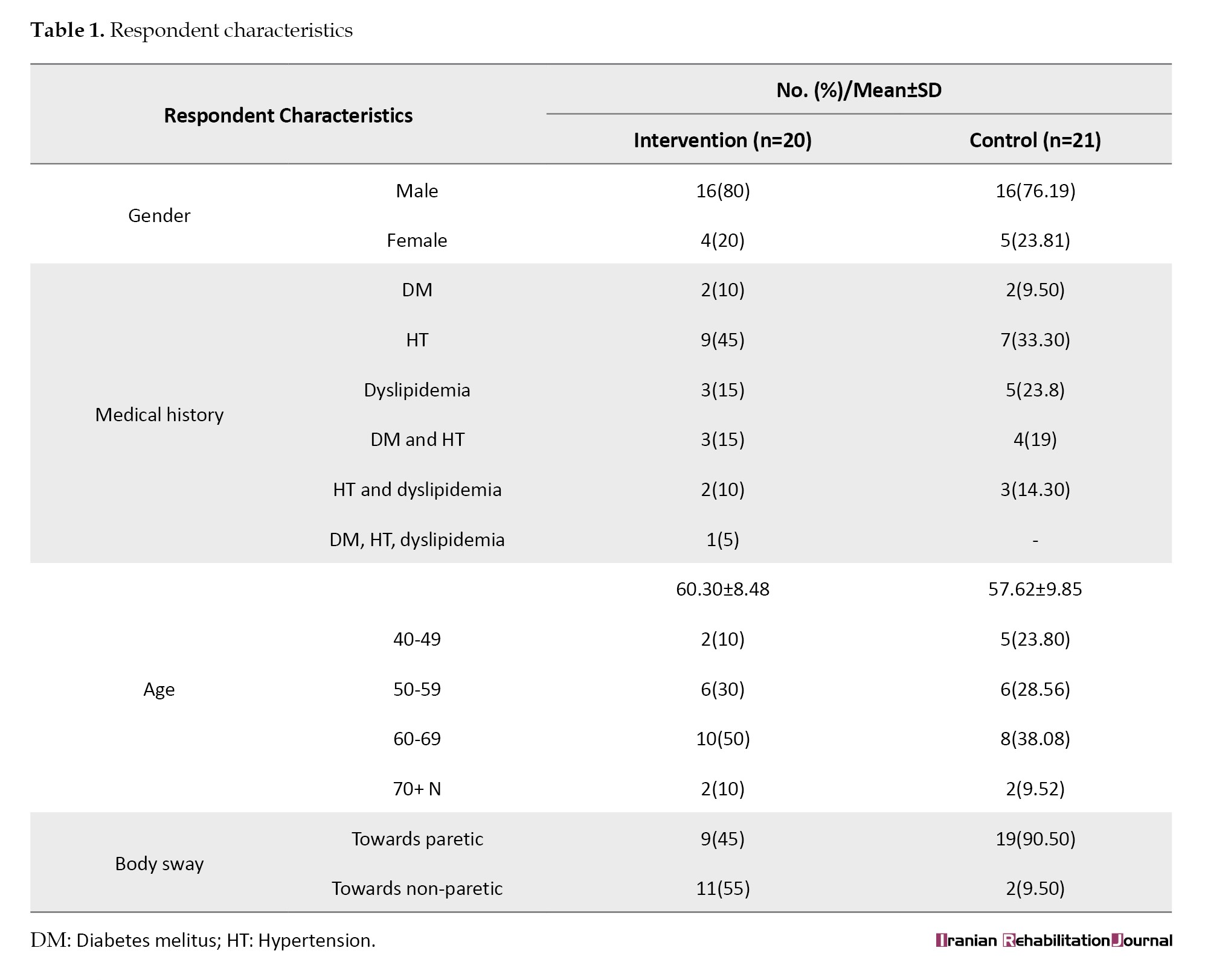
The intervention and control groups showed comparable distributions in terms of sex, age, medical history, and body sway direction, indicating similar baseline characteristics between the two groups. The normality and homogeneity test results are presented in Table 2.
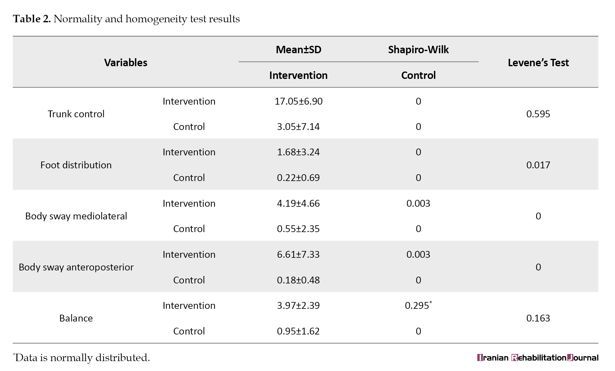
Data normality was assessed using the Shapiro–Wilk test, followed by Levene's Test to evaluate the homogeneity of variance between the intervention and control groups.
The Shapiro-Wilk test results showed that all variables in the intervention and control groups, such as trunk control, foot support distribution, mediolateral and anteroposterior body sway, show abnormal results (P<0.05) except for the balance variable in the intervention group.
The homogeneity test using Levene’s test showed that most variables had a significance value <0.05, particularly foot support distribution, mediolateral body sway, and anteroposterior body sway, which means that the data on these variables were not homogeneous between groups. Conversely, the trunk control and balance variables had a significance value >0.05; therefore, data on these variables were declared homogeneous.
The effect of postural training on postural stability in acute stroke
Determining the effect of postural training on postural stability using several measurements, namely the TCT to analyze trunk control, PBF to observe body sway and foot distribution, and the FRT to measure balance. The results of the measurements show the effect of postural training on postural stability in acute stroke in the intervention and control groups.
The results of the analysis showed that the intervention group experienced a significant increase in trunk control, body sway (mediolateral and anteroposterior), and balance before and after the intervention (P<0.05). In contrast, the control group only showed a significant effect on the balance variable. Furthermore, the foot distribution variable did not show a significant effect in either the intervention or control groups.
The effect of intergroup postural training on postural stability in acute stroke
Hypothesis testing was performed on the mediolateral body sway, anterolateral body sway, and mediolateral body sway variables using the Mann-Whitney test and the foot distribution and trunk control variables. Mann-Whitney analysis was used to compare the differences between the intervention and control groups on various postural stability parameters.
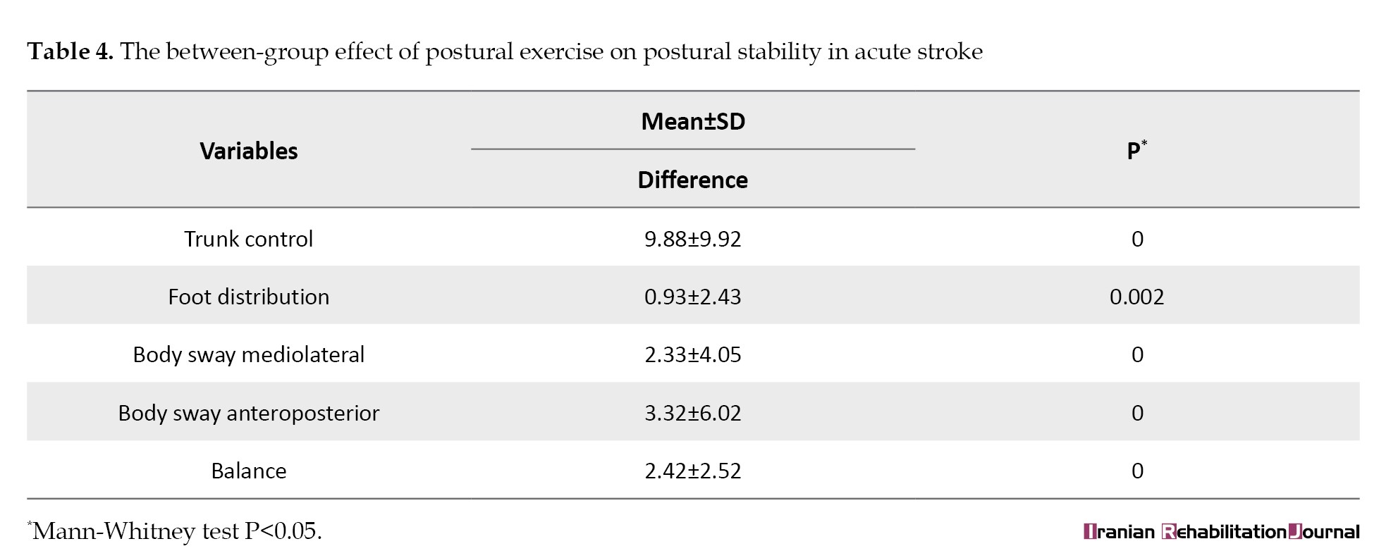
The results of the analysis showed significant differences between the values of all variables tested. Specifically, significant differences were observed between the two groups tested in terms of trunk control, foot distribution, mediolateral body sway, anteroposterior body sway, and balance. The intervention group demonstrated significant improvements in trunk control, mediolateral body sway, anteroposterior body sway, and functional balance after postural training with PBF (Table 3).
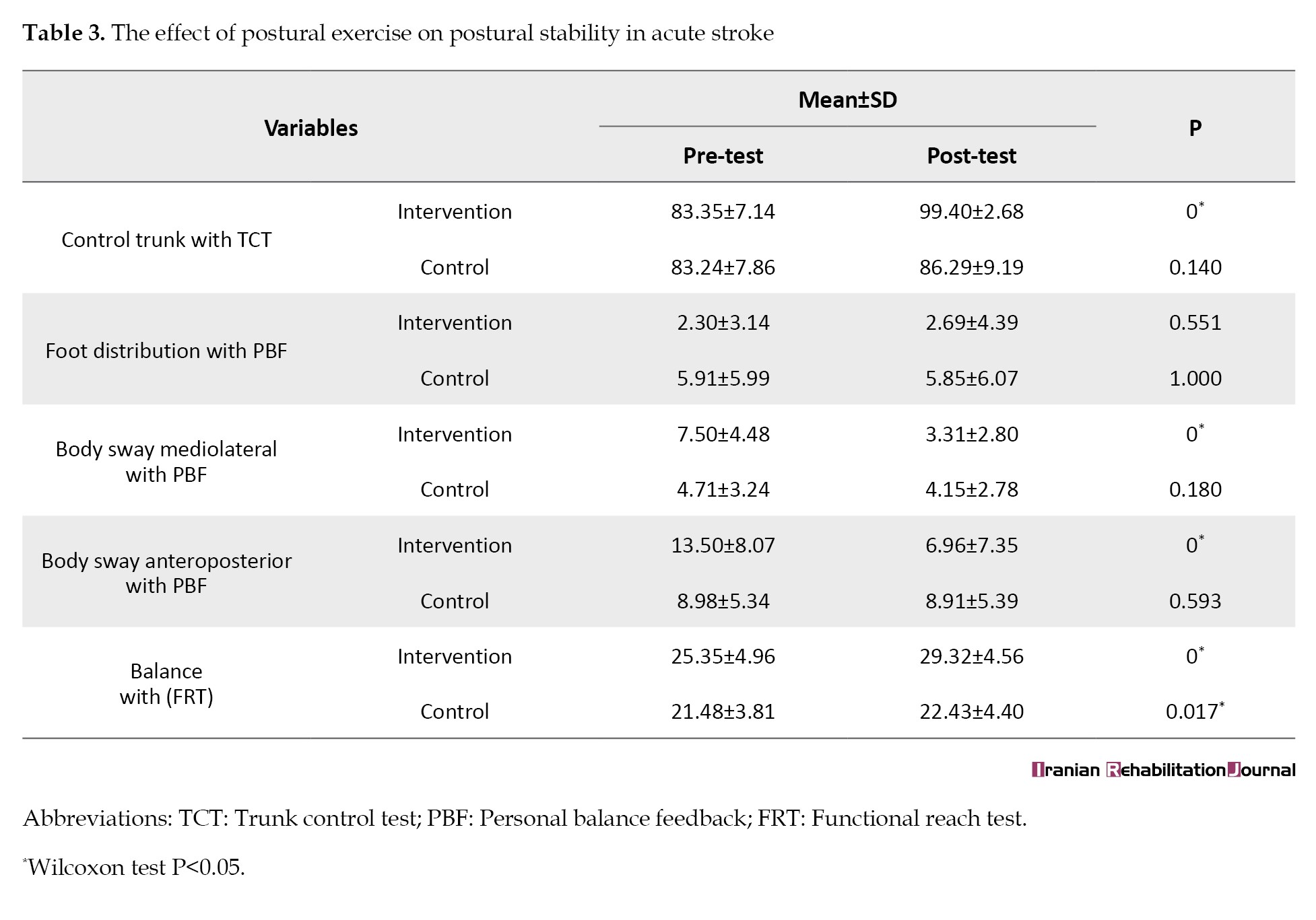
In contrast, the control group showed a significant improvement only in functional balance, while the other outcome measures remained unchanged. Between-group analysis revealed significant differences in trunk control, foot pressure distribution, mediolateral body sway, anteroposterior body sway, and functional balance, favoring the PBF intervention (Table 4).
Range of motion of the joint on the non-paretic side will increase. Meanwhile, in stroke patients who experience body sway towards the non-paretic side, this is due to spatial neglect, where patients experience distortion in the coordinates that are normally used to distribute this is because when is movement in the joint the joint will experience muscle contraction and increase somatosensory body weight on both legs when standing [37]. In some patients, the mid-sagittal plane used as a postural reference may shift to the side of the lesion and undergo contralateral rotation in the long body axis, resulting in body sway to the non-paretic side [38].
Discussion
The principal finding of this randomized controlled trial was that postural training using PBF significantly improved postural stability in patients with acute stroke compared with standard training, with a predominance of men (78%) over women (22%). This gender composition is commonly found in studies of elderly populations related to hypertension and chronic diseases. For example, the Sleman HDSS (Yogyakarta) study showed that hypertension was more common in men (42%) than women (38%). In addition, global data also show a slightly higher prevalence of high blood pressure in men than in women before the age of 50–60 years [32].
The largest age group was 60–69 years (34.1%), followed by 50–59 years (29.3%), 70+ (22%), and 40–49 years (14.6%). This is consistent with the trend of increasing stroke prevalence, in which age is associated with stroke incidence [33]. In older adults, postural stability declines, with a gradual decrease in the strength of the ankle stabilizing muscles, leading to dependence on the hip strategy with delayed activation of the trunk and thigh muscles, especially when performing activities and provoking a stable upright posture [34].
Most respondents had a history of hypertension (70.7%), while diabetes mellitus (29.3%) and dyslipidemia (34.1%) were also high. Hypertension is a risk factor for cerebrovascular disease, the leading cause of death [35]. Dyslipidemia plays a complex role in cerebrovascular disease, which has a strong association between cholesterol and ischemic stroke [33].
Stroke patients in this study experienced more body sway toward the paretic side (n=28). Stroke patients often experience muscle weakness on the paretic side due to damage to the corticospinal tract, which interferes with motor unit recruitment. This condition reduces the ability of the paretic limb to bear weight, causing the patient’s body weight to shift the CoM to the non-paretic side as compensation. Stroke patients who experience muscle weakness on one side will experience body sway toward the paretic side for reasons of safety and speed [36]. This is because when there is movement in the joint, the joint will experience muscle contraction and increase somatosensory input and muscular input, so that the range of motion of the joint on the non-paretic side will increase. Meanwhile, in stroke patients who experience body sway towards the non-paretic side, this is due to spatial neglect, where patients experience distortion in the coordinates that are normally used to distribute body weight on both legs when standing [37]. In some patients, the mid-sagittal plane used as a postural reference may shift to the side of the lesion and undergo contralateral rotation in the long body axis, resulting in body sway to the non-paretic side [38].
The effect of postural training with PBF on postural stability in acute stroke. The intervention group experienced a very significant increase from a mean value of 83.35±7.14 at baseline to 99.40±2.68 at the final measurement (P=0.000), while in the control group, the increase from 83.24±7.86 to 86.29±9.19 was not significant (P=0.140). Physiologically, this increase is closely related to the involvement of major postural muscles, such as the erector spinae, obliquus abdominis, and rectus abdominis muscles, which play a role in maintaining body balance and trunk stability [39]. Task-oriented postural exercises can increase neuromuscular activation and strengthen trunk control through the principles of neuroplasticity, such as visual feedback [40]. Exercises using PBF help patients integrate visual and proprioceptive sensory input simultaneously, accelerating neuromuscular responses to body position.
A study by [28] supports these findings, in which visual feedback training showed significant improvements in trunk muscle activation and balance in patients with hemiplegic stroke. Other studies have also shown that postural training can improve trunk control in stroke, which is consistent with this study [3, 13, 41]. The trunk ensures that the center of pressure remains above the BOS, allowing the body to maintain balance and reduce body sway. Impaired trunk control is associated with delayed motor development and reduced functional mobility [12, 41]. In conditions such as stroke or other neurological disorders, trunk control is often impaired, affecting the patient’s posture, balance, and functional abilities.
In the distribution of foot pressure measured with PBF, neither the intervention nor the control group showed significant changes, with the intervention group increasing slightly from 2.30±3.14 to 2.69±4.39 (P=0.551) and the control group decreasing slightly from 5.91±5.99 to 5.85±6.07 (P=1.000). This indicates that biomechanical changes, such as symmetry of weight bearing, require a longer training duration. According to [8]. Six weeks of training are needed to change the weight distribution pattern after a stroke. In the physiological distribution of weight on the feet after a stroke, patients tend to develop compensatory strategies by distributing their body weight more predominantly on the non-paretic side [34]. This is due to muscle weakness, sensory disturbances, and decreased body perception on the paretic side, so that the motor system tends to maintain this compensatory pattern to avoid the risk of falling. According to [42], asymmetry in weight distribution is a common condition after a stroke.
Foot pressure distribution is highly dependent on the integrity of the proprioceptive system and body awareness, which are often impaired after a stroke [43]. Although visual feedback can help strengthen visual control, proprioceptive disturbances on the paretic side make it difficult for patients to internally correct their weight distribution [44]. In the intervention group, despite the use of visual feedback, each individual’s neurocognitive response varied. Visual feedback requires good cognitive capacity and visual attention, which not all stroke patients may have, especially those with working memory impairment or attention deficits [45].
This can hinder the effectiveness of interventions in some participants. Significant changes in foot distribution require not only shifting exercises, but also leg muscle strengthening exercises, dynamic balance exercises, and the integration of adaptive postural strategies. If the intervention focuses only on shifting, even with feedback, the results may be limited [46].
The mediolateral body sway variable showed a significant decrease in the intervention group from 7.50±4.48 to 3.31±2.80 (P=0.000), while the control group experienced a non-significant decrease from 4.71±3.24 to 4.15±2.78 (P=0.180). In anteroposterior body sway, the intervention group also experienced a significant decrease from 13.50±8.07 to 6.96±7.35 (P=0.000), while the control group experienced a slight, insignificant decrease from 8.98±5.34 to 8.91±5.39 (P=0.593), indicating an improvement in the ability to maintain body tilt above the COG. This indicates that body sway is related to improved proprioceptive function and strengthening of the stabilizing muscles in the hips and trunk. The trunk functions as a central segment that helps maintain the body’s COG in the BOS [44]. Body sway is the movement of maintaining the body when the trunk rolls and tilts, which is a measure of postural stability to distinguish individuals with balance disorders or balanced body control [12]. Trunk movement control is necessary to maintain body balance and achieve smooth movements. Trunk control is a major contributor to balance and functional mobility [47].
This shows that exercises with visual feedback can actively improve postural stability. Body sway is related to natural movement and occurs when a person tries to maintain an upright posture while standing or performing activities [19]. This movement represents the body’s adjustment to maintain the COM within the BOS to prevent falling.
Previous studies have shown that postural training can change right and backward body sway [44]. These findings are consistent with a study by [48], which stated that dynamic balance training significantly reduced body sway, particularly in the mediolateral direction, which is most commonly impaired after stroke. This is important because excessive sway is closely associated with the risk of falling [49].
Meanwhile, in the balance variable measured by FRT, the intervention group experienced a significant increase from 25.35±4.96 to 29.32±4.56 (P=0.000), while the control group also increased from 21.48±3.81 to 22.43±4.40 with lower significance (P=0.017). Visual, vestibular, and somatosensory inputs contribute to maintaining postural stability by minimizing excessive sway [21]. Signals from the vestibular system help regulate the contraction of postural muscles, especially the trunk and limb muscles, so that the body can make automatic adjustments to maintain balance and reduce excessive body sway [21, 50]. The vestibular system integrates input from the eyes and proprioceptors (sensors of body and joint position) to produce an accurate perception of the body’s position in space [51, 52]. The results of the FRT showed a significant improvement (P=0.000) in the intervention group. This is consistent with previous studies [21, 53–56], which show that visual feedback can improve stability limits and functional abilities more quickly in both chronic and acute patients. This study has several limitations that should be considered when interpreting the findings. First, the intervention duration was relatively short (four days), and therefore, the long-term effects of PBF training remain unknown. Future studies should include follow-up assessments at 1 and 3 months post-intervention. Second, the sample size was relatively small (n=41), which may limit the statistical power to detect differences in secondary outcomes. Larger multicenter trials are needed to confirm these findings. Third, patients with cerebellar or brainstem strokes were excluded, limiting the generalizability of the results to these populations. Finally, some acute stroke patients were unable to tolerate standing balance training due to fatigue or severe motor impairment, which may introduce selection bias toward individuals with better early functional capacity.
Conclusion
Postural training using PBF appears to be effective in improving trunk control, reducing body sway, and enhancing functional balance in patients with acute stroke over a short-term period. However, further research with larger sample sizes and longer follow-up periods is required to determine the sustainability of these effects.
Ethical Considerations
Compliance with ethical guidelines
This study was approved by the Ethics Committee of Rumah Sakit Umum Pusat Dr. Sardjito (Dr. Sardjito General Hospital) Universitas Gajah Mada, Yogyakarta, Indonesia (Code: KE/FK/0390/EC/2025). All participants provided informed consent before data collection.
Funding
This research was a part of the research project and financially supported by the Research and Innovation Institute, Universitas Muhammadiyah Surakarta, Indonesia (Grant No.: 302.57/A.3-III/LRI/VIII/2024).
Authors' contributions
Conceptualization: Umi Budi Rahayu and Wanda Kurnia Yuda; Methodology: Wanda Kurnia Yuda, Umi Budi Rahayu, and Dwi Rosella Komalasari; Investigation and data collection: Wanda Kurnia Yuda; Formal analysis: Wanda Kurnia Yuda and Umi Budi Rahayu; Writing the original draft: Wanda Kurnia Yuda; Review and editing: Umi Budi Rahayu and Dwi Rosella Komalasari; Supervision: Umi Budi Rahayu; All authors read and approved the final manuscript.
Conflict of interest
The authors declared no conflicts of interest.
Acknowledgments
The authors thank the participants in this study, chairman of the Pusat Otak Nasional Hospital, research and Innovation Institute, Muhammadiyah University of Surakarta, and the Council for Higher Education, Research and Development, Muhammadiyah Organization for providing funding for this research.
Stroke is a clinical cerebrovascular disease with high morbidity, mortality, and disability rates, and has a heavy economic impact on families and communities [1]. Stroke also impairs neurological function due to blockage or rupture of blood vessels flowing to the brain [2, 3].
Blockage or rupture of blood vessels flowing to the brain results in decreased neurological function [4]. Patients will experience motor disorders such as loss of muscle control, movement, or mobility [5]. Stroke survivors usually experience long-term difficulties in performing daily activities and limitations in social life [6] and an inability to control posture, resulting in a high risk of falls and decreased postural stability [7].
Postural stability is the ability to shift weight onto paralyzed lower limbs. It is a prerequisite for functional mobility skills, such as rising from a chair, reaching while standing, and walking [8]. Perceptual dysfunction, sensory loss, poor motor recovery in the lower limbs, spasticity, muscle weakness, and decreased trunk control [9].
Trunk control is a major predictor of functional outcomes and hospital stay after stroke [10]. Trunk control acts as the central segment that helps maintain the center of gravity (COG) as the base of support (BOS) or the foundation of the body [11, 12]. The BOS, which is the foundation that supports the body, must remain at the COG [13]. The Trunk control test (TCT), which has a validity coefficient of 0.76, is used to assess trunk control [14].
Body sway plays an active role in determining postural stability [12]. Increased body sway reflects poorer postural stability and means that the body is less able to maintain balance effectively [1]. Body sway is also influenced by the vestibular system, which integrates input from the eyes and proprioceptors (sensors of body and joint position) to produce an accurate perception of the body’s position in space [9].
In stroke, there is an imbalance in the distribution of foot pressure or body weight between the affected (paretic) and unaffected (non-paretic) limbs [15]. Stroke patients tend to put more weight on their healthy leg. This imbalance in weight distribution makes it difficult to stabilize the center of mass (COM) above the BOS [16]. This disorder causes patients to rely on the healthy side of their body, resulting in the weaker side being less involved in maintaining balance and postural coordination [12]. The distribution of weight bearing also affects the vestibular system, namely, balance [9]. Postural stabilization refers to the ability to maintain body balance [17].
Balance is crucial for maintaining stability and controlling body movements [3, 18]. Balance refers to the ability to maintain the COM above the BOS [19]. The balance measurement tool for stroke is the functional reach test (FRT), which has a validity score of 0.71 [20]. Balance depends on the integration of sensory and motor systems by the central nervous system, which assigns different weights to sensory input from the visual, somatosensory, and vestibular or balance systems [19].
Stroke causes common balance problems that increase the risk of falls [21]. A total of 83% of patients with acute stroke develop postural imbalance and an increased risk of falling, ranging from 7% in the first week after stroke to 73% in the first year after discharge from hospital [7]. Therefore, exercises that target postural stabilization, such as postural exercises, are recommended [18, 22].
Postural exercises target improving postural stability by better controlling body sway [17, 23]. Exercises aimed at maintaining body posture, especially when performing postural exercises that lead to improved postural stabilization [24].
Previous studies have shown that certain exercises for postural stabilization using robots can promote rapid recovery of balance and motor function [25–27]. Exercises using visual feedback can improve trunk stability in patients with stroke [21, 28–30]. One tool that can be used to improve postural stabilization is personal balance feedback (PBF). PBF has been registered as a simple patent with patent number IDS00001023. PBF is a tool consisting of scales and a smart belt. Patients are instructed to stand on the device and wear a smart belt. This will display the patient’s body sway and weight distribution on the monitor screen in real-time. Patients are instructed to straighten their bodies using the monitor screen.
Previous studies related to interventions that can improve postural stability in stroke patients have been conducted on patients with chronic stroke [8, 19, 26, 28, 31]. Therefore, there are still limitations in studies discussing postural stability interventions in acute stroke patients. Therefore, this study focuses on postural control exercises in acute stroke so that it can be used as a reference for providing appropriate interventions in the acute phase.
Materials and Methods
Design and participants
This was a randomized controlled trial conducted at Rumah Sakit Pusat Otak Nasional Prof. Dr. dr. Mahar Mardjono (RS PON)/Prof. Dr. dr. Mahar Mardjono National Brain Center Hospital. A total of 41 patients with acute stroke (≤48 hours) who met the inclusion criteria were randomly assigned to the intervention and control groups. Participants in the intervention group performed postural exercises using PBF while standing with both feet shoulder-width apart on a smart scale and wearing a smart belt. A walker was provided in front of the patient to prevent falls. Real-time visual feedback on foot pressure distribution and body tilt was displayed on a monitor in front of the participant. The exercise protocol consisted of anteroposterior body sway, mediolateral body sway, and foot weight distribution. Participants were instructed to shift their weight to the paretic side while observing the visual feedback. The duration of each session was gradually increased based on the principles of exercise overload and neural rehabilitation: 5 minutes on day 1, 10 minutes on day 2, 15 minutes on day 3, and 20 minutes on day 4. This progression was adapted from previous early mobilization protocols in acute stroke rehabilitation.
The control group received standard weight shift training. Evaluations were conducted before and after the intervention using TCT, FRT, and data from PBF for body sway (anteroposterior and mediolateral) and foot distribution. Statistical analysis was performed using the Mann-Whitney test and independent t-test with a significance level of P<0.05.
Data collection
Participants were recruited from the inpatient ward of the RS PON in Jakarta based on inclusion and exclusion criteria. The inclusion criteria were patients with a diagnosis of ischemic stroke confirmed by a neurologist, patients with stable hemodynamics confirmed by the attending physician, patients who were conscious and cooperative in communication, patients who were able to sit on the second day after stroke onset, and patients who had a minimum motor strength of 3. The exclusion criteria were infarcts in the cerebellum and brainstem, joint deformities, and contractures affecting gait and balance. The dropout criterion was the inability to stand on the third day after stroke onset.
Participants were randomly allocated to the intervention or control group using a computer-generated random sequence created by an independent statistician who was not involved in participant recruitment or outcome assessment. A simple randomization method with a 1:1 allocation ratio was applied. Allocation concealment was ensured using an online spinner with numbers 1 and 2, where 1 was the intervention group and 2 was the control group. This procedure minimized selection bias and ensured that the researchers responsible for recruitment did not know the group allocation prior to assignment.
Data and information selected based on characteristics related to inclusion were processed and analyzed objectively and systematically from the data obtained. Trunk control was assessed using the TCT, demonstrating good construct validity (r=0.76) and excellent inter-rater reliability (intraclass correlation coefficient [ICC]>0.90) in stroke populations, which consists of four items: Rolling to the healthy side, rolling to the weak side, sitting up from a lying position, and maintaining a sitting position for 30 seconds. Functional balance was assessed using the FRT, widely used in stroke rehabilitation, and showed acceptable validity (r=0.71) and high test-re-test reliability (ICC>0.85) by measuring the maximum distance that participants can reach forward without losing balance. Body sway (anteroposterior and mediolateral) and right-left foot distribution were measured using the PBF system, which was previously tested for measurement consistency in pilot studies, demonstrating acceptable agreement, which provided a visualization of the participant’s body weight while standing and body sway.
Results
Data normality was assessed using the Shapiro-Wilk test. As most variables did not follow a normal distribution, non-parametric tests were applied. Within-group comparisons were analyzed using the Wilcoxon signed-rank test, while between-group differences were assessed using the Mann–Whitney U test. Effect sizes were calculated using r values to determine the magnitude of the intervention effect. Statistical significance was set at P<0.05.
The characteristics of respondents in the intervention and control groups showed a balanced demographic and clinical distribution. Most respondents were male in both groups (intervention: 80%; control: 76.19%). The average age was similar between the intervention group (60.30±8.48 years) and the control group (57.62±9.85 years). The most common medical history was hypertension (intervention: 45%; control: 33.3%). The distribution of body sway direction showed a striking difference, where the majority of respondents in the control group experienced a shift towards the paretic side (90.5%), while in the intervention group, it was more evenly distributed between paretic (45%) and non-paretic (55%).
The baseline demographic and clinical characteristics of the participants are presented in Table 1.
The intervention and control groups showed comparable distributions in terms of sex, age, medical history, and body sway direction, indicating similar baseline characteristics between the two groups. The normality and homogeneity test results are presented in Table 2.
Data normality was assessed using the Shapiro–Wilk test, followed by Levene's Test to evaluate the homogeneity of variance between the intervention and control groups.
The Shapiro-Wilk test results showed that all variables in the intervention and control groups, such as trunk control, foot support distribution, mediolateral and anteroposterior body sway, show abnormal results (P<0.05) except for the balance variable in the intervention group.
The homogeneity test using Levene’s test showed that most variables had a significance value <0.05, particularly foot support distribution, mediolateral body sway, and anteroposterior body sway, which means that the data on these variables were not homogeneous between groups. Conversely, the trunk control and balance variables had a significance value >0.05; therefore, data on these variables were declared homogeneous.
The effect of postural training on postural stability in acute stroke
Determining the effect of postural training on postural stability using several measurements, namely the TCT to analyze trunk control, PBF to observe body sway and foot distribution, and the FRT to measure balance. The results of the measurements show the effect of postural training on postural stability in acute stroke in the intervention and control groups.
The results of the analysis showed that the intervention group experienced a significant increase in trunk control, body sway (mediolateral and anteroposterior), and balance before and after the intervention (P<0.05). In contrast, the control group only showed a significant effect on the balance variable. Furthermore, the foot distribution variable did not show a significant effect in either the intervention or control groups.
The effect of intergroup postural training on postural stability in acute stroke
Hypothesis testing was performed on the mediolateral body sway, anterolateral body sway, and mediolateral body sway variables using the Mann-Whitney test and the foot distribution and trunk control variables. Mann-Whitney analysis was used to compare the differences between the intervention and control groups on various postural stability parameters.
The results of the analysis showed significant differences between the values of all variables tested. Specifically, significant differences were observed between the two groups tested in terms of trunk control, foot distribution, mediolateral body sway, anteroposterior body sway, and balance. The intervention group demonstrated significant improvements in trunk control, mediolateral body sway, anteroposterior body sway, and functional balance after postural training with PBF (Table 3).
In contrast, the control group showed a significant improvement only in functional balance, while the other outcome measures remained unchanged. Between-group analysis revealed significant differences in trunk control, foot pressure distribution, mediolateral body sway, anteroposterior body sway, and functional balance, favoring the PBF intervention (Table 4).
Range of motion of the joint on the non-paretic side will increase. Meanwhile, in stroke patients who experience body sway towards the non-paretic side, this is due to spatial neglect, where patients experience distortion in the coordinates that are normally used to distribute this is because when is movement in the joint the joint will experience muscle contraction and increase somatosensory body weight on both legs when standing [37]. In some patients, the mid-sagittal plane used as a postural reference may shift to the side of the lesion and undergo contralateral rotation in the long body axis, resulting in body sway to the non-paretic side [38].
Discussion
The principal finding of this randomized controlled trial was that postural training using PBF significantly improved postural stability in patients with acute stroke compared with standard training, with a predominance of men (78%) over women (22%). This gender composition is commonly found in studies of elderly populations related to hypertension and chronic diseases. For example, the Sleman HDSS (Yogyakarta) study showed that hypertension was more common in men (42%) than women (38%). In addition, global data also show a slightly higher prevalence of high blood pressure in men than in women before the age of 50–60 years [32].
The largest age group was 60–69 years (34.1%), followed by 50–59 years (29.3%), 70+ (22%), and 40–49 years (14.6%). This is consistent with the trend of increasing stroke prevalence, in which age is associated with stroke incidence [33]. In older adults, postural stability declines, with a gradual decrease in the strength of the ankle stabilizing muscles, leading to dependence on the hip strategy with delayed activation of the trunk and thigh muscles, especially when performing activities and provoking a stable upright posture [34].
Most respondents had a history of hypertension (70.7%), while diabetes mellitus (29.3%) and dyslipidemia (34.1%) were also high. Hypertension is a risk factor for cerebrovascular disease, the leading cause of death [35]. Dyslipidemia plays a complex role in cerebrovascular disease, which has a strong association between cholesterol and ischemic stroke [33].
Stroke patients in this study experienced more body sway toward the paretic side (n=28). Stroke patients often experience muscle weakness on the paretic side due to damage to the corticospinal tract, which interferes with motor unit recruitment. This condition reduces the ability of the paretic limb to bear weight, causing the patient’s body weight to shift the CoM to the non-paretic side as compensation. Stroke patients who experience muscle weakness on one side will experience body sway toward the paretic side for reasons of safety and speed [36]. This is because when there is movement in the joint, the joint will experience muscle contraction and increase somatosensory input and muscular input, so that the range of motion of the joint on the non-paretic side will increase. Meanwhile, in stroke patients who experience body sway towards the non-paretic side, this is due to spatial neglect, where patients experience distortion in the coordinates that are normally used to distribute body weight on both legs when standing [37]. In some patients, the mid-sagittal plane used as a postural reference may shift to the side of the lesion and undergo contralateral rotation in the long body axis, resulting in body sway to the non-paretic side [38].
The effect of postural training with PBF on postural stability in acute stroke. The intervention group experienced a very significant increase from a mean value of 83.35±7.14 at baseline to 99.40±2.68 at the final measurement (P=0.000), while in the control group, the increase from 83.24±7.86 to 86.29±9.19 was not significant (P=0.140). Physiologically, this increase is closely related to the involvement of major postural muscles, such as the erector spinae, obliquus abdominis, and rectus abdominis muscles, which play a role in maintaining body balance and trunk stability [39]. Task-oriented postural exercises can increase neuromuscular activation and strengthen trunk control through the principles of neuroplasticity, such as visual feedback [40]. Exercises using PBF help patients integrate visual and proprioceptive sensory input simultaneously, accelerating neuromuscular responses to body position.
A study by [28] supports these findings, in which visual feedback training showed significant improvements in trunk muscle activation and balance in patients with hemiplegic stroke. Other studies have also shown that postural training can improve trunk control in stroke, which is consistent with this study [3, 13, 41]. The trunk ensures that the center of pressure remains above the BOS, allowing the body to maintain balance and reduce body sway. Impaired trunk control is associated with delayed motor development and reduced functional mobility [12, 41]. In conditions such as stroke or other neurological disorders, trunk control is often impaired, affecting the patient’s posture, balance, and functional abilities.
In the distribution of foot pressure measured with PBF, neither the intervention nor the control group showed significant changes, with the intervention group increasing slightly from 2.30±3.14 to 2.69±4.39 (P=0.551) and the control group decreasing slightly from 5.91±5.99 to 5.85±6.07 (P=1.000). This indicates that biomechanical changes, such as symmetry of weight bearing, require a longer training duration. According to [8]. Six weeks of training are needed to change the weight distribution pattern after a stroke. In the physiological distribution of weight on the feet after a stroke, patients tend to develop compensatory strategies by distributing their body weight more predominantly on the non-paretic side [34]. This is due to muscle weakness, sensory disturbances, and decreased body perception on the paretic side, so that the motor system tends to maintain this compensatory pattern to avoid the risk of falling. According to [42], asymmetry in weight distribution is a common condition after a stroke.
Foot pressure distribution is highly dependent on the integrity of the proprioceptive system and body awareness, which are often impaired after a stroke [43]. Although visual feedback can help strengthen visual control, proprioceptive disturbances on the paretic side make it difficult for patients to internally correct their weight distribution [44]. In the intervention group, despite the use of visual feedback, each individual’s neurocognitive response varied. Visual feedback requires good cognitive capacity and visual attention, which not all stroke patients may have, especially those with working memory impairment or attention deficits [45].
This can hinder the effectiveness of interventions in some participants. Significant changes in foot distribution require not only shifting exercises, but also leg muscle strengthening exercises, dynamic balance exercises, and the integration of adaptive postural strategies. If the intervention focuses only on shifting, even with feedback, the results may be limited [46].
The mediolateral body sway variable showed a significant decrease in the intervention group from 7.50±4.48 to 3.31±2.80 (P=0.000), while the control group experienced a non-significant decrease from 4.71±3.24 to 4.15±2.78 (P=0.180). In anteroposterior body sway, the intervention group also experienced a significant decrease from 13.50±8.07 to 6.96±7.35 (P=0.000), while the control group experienced a slight, insignificant decrease from 8.98±5.34 to 8.91±5.39 (P=0.593), indicating an improvement in the ability to maintain body tilt above the COG. This indicates that body sway is related to improved proprioceptive function and strengthening of the stabilizing muscles in the hips and trunk. The trunk functions as a central segment that helps maintain the body’s COG in the BOS [44]. Body sway is the movement of maintaining the body when the trunk rolls and tilts, which is a measure of postural stability to distinguish individuals with balance disorders or balanced body control [12]. Trunk movement control is necessary to maintain body balance and achieve smooth movements. Trunk control is a major contributor to balance and functional mobility [47].
This shows that exercises with visual feedback can actively improve postural stability. Body sway is related to natural movement and occurs when a person tries to maintain an upright posture while standing or performing activities [19]. This movement represents the body’s adjustment to maintain the COM within the BOS to prevent falling.
Previous studies have shown that postural training can change right and backward body sway [44]. These findings are consistent with a study by [48], which stated that dynamic balance training significantly reduced body sway, particularly in the mediolateral direction, which is most commonly impaired after stroke. This is important because excessive sway is closely associated with the risk of falling [49].
Meanwhile, in the balance variable measured by FRT, the intervention group experienced a significant increase from 25.35±4.96 to 29.32±4.56 (P=0.000), while the control group also increased from 21.48±3.81 to 22.43±4.40 with lower significance (P=0.017). Visual, vestibular, and somatosensory inputs contribute to maintaining postural stability by minimizing excessive sway [21]. Signals from the vestibular system help regulate the contraction of postural muscles, especially the trunk and limb muscles, so that the body can make automatic adjustments to maintain balance and reduce excessive body sway [21, 50]. The vestibular system integrates input from the eyes and proprioceptors (sensors of body and joint position) to produce an accurate perception of the body’s position in space [51, 52]. The results of the FRT showed a significant improvement (P=0.000) in the intervention group. This is consistent with previous studies [21, 53–56], which show that visual feedback can improve stability limits and functional abilities more quickly in both chronic and acute patients. This study has several limitations that should be considered when interpreting the findings. First, the intervention duration was relatively short (four days), and therefore, the long-term effects of PBF training remain unknown. Future studies should include follow-up assessments at 1 and 3 months post-intervention. Second, the sample size was relatively small (n=41), which may limit the statistical power to detect differences in secondary outcomes. Larger multicenter trials are needed to confirm these findings. Third, patients with cerebellar or brainstem strokes were excluded, limiting the generalizability of the results to these populations. Finally, some acute stroke patients were unable to tolerate standing balance training due to fatigue or severe motor impairment, which may introduce selection bias toward individuals with better early functional capacity.
Conclusion
Postural training using PBF appears to be effective in improving trunk control, reducing body sway, and enhancing functional balance in patients with acute stroke over a short-term period. However, further research with larger sample sizes and longer follow-up periods is required to determine the sustainability of these effects.
Ethical Considerations
Compliance with ethical guidelines
This study was approved by the Ethics Committee of Rumah Sakit Umum Pusat Dr. Sardjito (Dr. Sardjito General Hospital) Universitas Gajah Mada, Yogyakarta, Indonesia (Code: KE/FK/0390/EC/2025). All participants provided informed consent before data collection.
Funding
This research was a part of the research project and financially supported by the Research and Innovation Institute, Universitas Muhammadiyah Surakarta, Indonesia (Grant No.: 302.57/A.3-III/LRI/VIII/2024).
Authors' contributions
Conceptualization: Umi Budi Rahayu and Wanda Kurnia Yuda; Methodology: Wanda Kurnia Yuda, Umi Budi Rahayu, and Dwi Rosella Komalasari; Investigation and data collection: Wanda Kurnia Yuda; Formal analysis: Wanda Kurnia Yuda and Umi Budi Rahayu; Writing the original draft: Wanda Kurnia Yuda; Review and editing: Umi Budi Rahayu and Dwi Rosella Komalasari; Supervision: Umi Budi Rahayu; All authors read and approved the final manuscript.
Conflict of interest
The authors declared no conflicts of interest.
Acknowledgments
The authors thank the participants in this study, chairman of the Pusat Otak Nasional Hospital, research and Innovation Institute, Muhammadiyah University of Surakarta, and the Council for Higher Education, Research and Development, Muhammadiyah Organization for providing funding for this research.
References
- GBD 2017 Disease and Injury Incidence and Prevalence Collaborators. Global, regional, and national incidence, prevalence, and years lived with disability for 354 diseases and injuries for 195 countries and territories, 1990-2017: A systematic analysis for the Global Burden of Disease Study 2017. The Lancet. 2018; 392(10159):1789-858. [DOI:10.1016/s0140-6736(18)32279-7] [PMID]
- Kuriakose D, Xiao Z. Pathophysiology and treatment of stroke: Present status and future perspectives. International Journal of Molecular Sciences. 2020; 21(20):7609. [DOI:10.3390/ijms21207609] [PMID]
- Yeo SS, Koo DK, Ko SY, Park SY. Effect of Balance Training in Sitting Position Using Visual Feedback on Balance and Gait Ability in Chronic Stroke Patients. Journal of Clinical Medicine. 2023; 12(13):4383. [DOI:10.3390/jcm12134383] [PMID]
- Khan F, Chevidikunnan MF. Prevalence of balance impairment and factors associated with balance among patients with stroke. A cross sectional retrospective case control study. Healthcare (Switzerland) 2021; 9(3):320. [DOI:10.3390/healthcare9030320] [PMID]
- Hugues A, Di Marco J, Janiaud P, Xue Y, Pires J, Khademi H, et al. Efficiency of physical therapy on postural imbalance after stroke: Study protocol for a systematic review and meta-analysis. BMJ Open 2017; 7(1):e013348. [DOI:10.1136/bmjopen-2016-013348] [PMID]
- Lando A, Cacciante L, Mantineo A, Baldan F, Pillastrini P, Turolla A, et al. Multi-Modal versus Uni-Modal Treatment for the Recovery of Lower Limb Motor Function in Patients after Stroke: A Systematic Review with Meta-Analysis. Healthcare (Switzerland) 2024; 12(2):189. [DOI:10.3390/healthcare12020189] [PMID]
- Djurovic O, Mihaljevic O, Radovanovic S, Kostic S, Vukicevic M, Brkic BG, et al. Risk factors related to falling in patients after stroke. Iranian Journal of Public Health. 2021; 50(9):1832-41. [DOI:10.18502/ijph.v50i9.7056] [PMID]
- Komiya M, Maeda N, Narahara T, Suzuki Y, Fukui K, Tsutsumi S, et al. Effect of 6-week balance exercise by real-time postural feedback system on walking ability for patients with chronic stroke: A pilot single-blind randomized controlled trial. Brain Sciences. 2021; 11(11):1493. [DOI:10.3390/brainsci11111493] [PMID]
- Duchene Y, Mornieux G, Petel A, Perrin PP, Gauchard GC. The trunk’s contribution to postural control under challenging balance conditions. Gait & Posture. 2021; 84:102-7. [PMID]
- Van Criekinge T, Truijen S, Schröder J, Maebe Z, Blanckaert K, van der Waal C, et al. The effectiveness of trunk training on trunk control, sitting and standing balance and mobility post-stroke: A systematic review and meta-analysis. Clinical Rehabilitation. 2019; 33(6):992-1002. [DOI:10.1177/0269215519830159] [PMID]
- Allum JH, Adkin AL, Carpenter MG, Held-Ziolkowska M, Honegger F, Pierchala K. Trunk sway measures of postural stability during clinical balance tests: Effects of a unilateral vestibular deficit. Gait & Posture. 2001; 14(3):227-37.[DOI:10.1016/S0966-6362(01)00132-1] [PMID]
- Ha SY, Sung YH. Vojta Therapy Affects Trunk Control and Postural Sway in Children with Central Hypotonia: A Randomized Controlled Trial. Children. 2022; 9(10):1470. [DOI:10.3390/children9101470] [PMID]
- Karthikbabu S, Verheyden G. Relationship between trunk control, core muscle strength and balance confidence in community-dwelling patients with chronic stroke. Topics in Stroke Rehabilitation. 2021; 28(2):88-95. [DOI:10.1080/10749357.2020.1783896] [PMID]
- Verheyden G, Nieuwboer A, Van de Winckel A, De Weerdt W. Clinical tools to measure trunk performance after stroke: A systematic review of the literature. Clinical Rehabilitation. 2007; 21(5):387-94. [DOI:10.1177/0269215507074055] [PMID]
- Inoue M, Amimoto K, Chiba Y, Sekine D, Fukata K, Fujino Y, et al. Effect of Exercise Involving Standing Weight Shifting to the Nonparetic Side on an Inclined Surface in the Early Phase after a Stroke: A Randomized Controlled Trial. Physical Therapy. 2021; 101(8):pzab114. [DOI:10.1093/ptj/pzab114] [PMID]
- Shrestha R, Sandesh TS, Jalal Z, Nuhmani S, Alghadir AH, Khan M. Effects of multi-directional step exercise with weight-shifting as an adjunct to conventional exercises on balance and gait in stroke patients. Scientific Reports. 2022; 12(1):17053. [DOI:10.1038/s41598-022-21073-y] [PMID]
- Kimijanová J, Svoboda Z, Han J. Editorial: Sensory control of posture and gait: Integration and mechanisms to maintain balance during different sensory conditions. Frontiers in Human Neuroscience. 2024; 18:1378599. [DOI:10.3389/fnhum.2024.1378599] [PMID]
- Surgent OJ, Dadalko OI, Pickett KA, Travers BG. Balance and the brain: A review of structural brain correlates of postural balance and balance training in humans. Gait & Posture. 2019; 71:245-52. [DOI:10.1016/j.gaitpost.2019.05.011] [PMID]
- Ghrouz A, Guillen-Sola A, Morgado-Perez A, Muñoz-Redondo E, Ramírez-Fuentes C, Curbelo Peña Y, et al. The effect of a motor relearning on balance and postural control in patients after stroke: An open-label randomized controlled trial. European Stroke Journal. 2024; 9(2):303-11. [DOI:10.1177/23969873231220218] [PMID]
- Outermans JC, van Peppen RP, Wittink H, Takken T, Kwakkel G. Effects of a high-intensity task-oriented training on gait performance early after stroke: A pilot study. Clinical Rehabilitation. 2010; 24(11):979-87. [DOI:10.1177/0269215509360647] [PMID]
- Firdaus HM, Rahayu UB, Maghfiroh RA. Visual Effects of Biofeedback on Post-stroke Patients’ Balance: A Literature Review. Paper presented at: International Conference on Health and Well-Being (ICHWB 2022). 5-6 December 2022; Surakarta, Central Java, Indonesia. [DOI:10.2991/978-94-6463-184-5_33]
- Hyun SJ, Lee J, Lee BH. The effects of sit-to-stand training combined with real-time visual feedback on strength, balance, gait ability, and quality of life in patients with stroke: A randomized controlled trial. International Journal of Environmental Research and Public Health. 2021; 18(22):12229. [DOI:10.3390/ijerph182212229] [PMID]
- Ghorbanpour Z, Taghizadeh G, Hosseini SA, Pishyareh E, Ghomsheh FT, Bakhshi E, et al. Overload of anxiety on postural control impairments in chronic stroke survivors: The role of external focus and cognitive task on the automaticity of postural control. PLoS One. 2021; 16(7):e0252131. [DOI:10.1371/journal.pone.0252131] [PMID]
- Alsubaie SF. The postural stability measures most related to aging, physical performance, and cognitive function in healthy adults. BioMed Research International. 2020; 2020:5301534. DOI:10.1155/2020/5301534] [PMID]
- De Luca A, Squeri V, Barone LM, Vernetti Mansin H, Ricci S, Pisu I, et al. Dynamic Stability and Trunk Control Improvements Following Robotic Balance and Core Stability Training in Chronic Stroke Survivors: A Pilot Study. Frontiers in Neurology. 2020; 11:494. [DOI:10.3389/fneur.2020.00494] [PMID]
- Inoue S, Otaka Y, Kumagai M, Sugasawa M, Mori N, Kondo K. Effects of Balance Exercise Assist Robot training for patients with hemiparetic stroke: A randomized controlled trial. Journal of Neuroengineering and Rehabilitation. 2022; 19(1):12. [DOI:10.1186/s12984-022-00989-6] [PMID]
- Zhang Y, Zhao W, Wan C, Wu X, Huang J, Wang X, et al. Exoskeleton rehabilitation robot training for balance and lower limb function in sub-acute stroke patients: A pilot, randomized controlled trial. Journal of Neuroengineering and Rehabilitation. 2024; 21(1):98. [DOI:10.1186/s12984-024-01391-0] [PMID]
- Pak NW, Lee JH. Effects of visual feedback training and visual targets on muscle activation, balancing, and walking ability in adults after hemiplegic stroke: A preliminary, randomized, controlled study. International Journal of Rehabilitation Research. 2020; 43(1):76-81. [DOI:10.1097/MRR.0000000000000376] [PMID]
- Pellegrino L, Giannoni P, Marinelli L, Casadio M. Effects of continuous visual feedback during sitting balance training in chronic stroke survivors. Journal of Neuroengineering and Rehabilitation. 2017; 14(1):107. [DOI:10.1186/s12984-017-0316-0] [PMID]
- Ahmed M, Elshinnawy, Fathy KA, Wadee AN, Fayed IH. Effect of Visual Biofeedback Training on Postural Instability in Chronic Stroke Patients: A Controlled Randomized Trial. International Journal of Pharmaceutical Research. 2021; 13(2):2052-61. [DOI:10.31838/ijpr/2021.13.02.273]
- Lyu T, Yan K, Lyu J, Zhao X, Wang R, Zhang C, et al. Comparative efficacy of gait training for balance outcomes in patients with stroke: A systematic review and network meta-analysis. Frontiers in Neurology. 2023; 14:1093779. [DOI:10.3389/fneur.2023.1093779] [PMID]
- Defianna SR, Santosa A, Probandari A, Dewi FST. Gender Differences in Prevalence and Risk Factors for Hypertension among Adult Populations: A Cross-Sectional Study in Indonesia. International Journal of Environmental Research and Public Health. 2021; 18(12):6259. [DOI:10.3390/ijerph18126259] [PMID]
- Juli C, Heryaman H, Arnengsih, Ang ET, Defi IR, Gamayani U, et al. The number of risk factors increases the recurrence events in ischemic stroke. European Journal of Medical Research. 2022; 27(1):138. [DOI:10.1186/s40001-022-00768-y] [PMID]
- Liang HW, Chi SY, Tai TL, Li YH, Hwang YH. Impact of age on the postural stability measured by a virtual reality tracker-based posturography and a pressure platform system. BMC Geriatrics. 2022; 22(1):506. [DOI:10.1186/s12877-022-03195-0] [PMID]
- Sethi R, Hiremath JS, Ganesh V, Banerjee S, Shah M, Mehta A, et al. Correlation between Stroke Risk and Systolic Blood Pressure in Patients over 50 Years with Uncontrolled Hypertension: Results from the SYSTUP-India Study. Cardiovascular Therapeutics. 2021; 2021:6622651. [DOI:10.1155/2021/6622651] [PMID]
- Gammeri R, Iacono C, Ricci R, Salatino A. Unilateral spatial neglect after stroke: Current insights. Neuropsychiatric Disease and Treatment. 2020; 16:131-52. [DOI:10.2147/NDT.S171461] [PMID]
- Williams LJ, Kernot J, Hillier SL, Loetscher T. Spatial Neglect Subtypes, Definitions and Assessment Tools: A Scoping Review. Frontiers in Neurology. 2021; 12:742365. [DOI:10.3389/fneur.2021.742365] [PMID]
- Li K, Malhotra PA. Spatial neglect. Practical Neurology. 2015; 15(5):333-9. [DOI:10.1136/practneurol-2015-001115] [PMID]
- Nakata A, Osuka S, Ishida T, Saito Y, Samukawa M, Kasahara S, et al. Trunk Muscle Activity and Ratio of Local Muscle to Global Muscle Activity during Supine Bridge Exercises under Unstable Conditions in Young Participants with and without Chronic Low Back Pain. Healthcare (Switzerland). 2024; 12(5):514. [DOI:10.3390/healthcare12050514] [PMID]
- Khallaf ME. Effect of Task-Specific Training on Trunk Control and Balance in Patients with Subacute Stroke. Neurology Research International. 2020; 2020:5090193.[DOI:10.1155/2020/5090193] [PMID]
- Appeadu MK, Gupta V. Postural instability. Treasure Island: StatPearls Publishing; 2026. [Link]
- Little VL, Perry LA, Mercado MWV, Kautz SA, Patten C. Gait asymmetry pattern following stroke determines acute response to locomotor task. Gait & Posture. 2020; 77:300-7.[DOI:10.1016/j.gaitpost.2020.02.016] [PMID]
- Song Q, Zhang X, Mao M, Sun W, Zhang C, Chen Y, et al. Relationship of proprioception, cutaneous sensitivity, and muscle strength with the balance control among older adults. Journal of Sport and Health Science. 2021; 10(5):585-93. [DOI:10.1016/j.jshs.2021.07.005] [PMID]
- Jo YJ, Kim DH, Kim S, Kim JH, Choi JH, Park JB, et al. Effect of Anterioposterior Weight-Shift Training with Visual Biofeedback in Patients with Step Length Asymmetry after Subacute Stroke. Journal of Personalized Medicine. 2023; 13(12):1726. [DOI:10.3390/jpm13121726] [PMID]
- Tai RY, Zhu JD, Chen CC, Hsieh YW, Cheng CH. Modulation of functional connectivity in response to mirror visual feedback in stroke survivors: An MEG study. Brain Sciences. 2021; 11(10):1284. [DOI:10.3390/brainsci11101284] [PMID]
- No SM, Hwang YT, Son SM. The effect of postural balance and fall efficacy on bilateral visual feedback training with visual targets in stroke patients. The Journal of Korean Physical Therapy. 2022; 34(2):57-62. [Link]
- Schöne B, Kisker J, Lange L, Gruber T, Sylvester S, Osinsky R. The reality of virtual reality. Frontiers in Psychology. 2023; 14:1093014. [DOI:10.3389/fpsyg.2023.1093014] [PMID]
- Soke F, Eldemir S, Ozkan T, Ozkul C, Ozcan Gulsen E, Gulsen C, et al. The functional reach test in people with multiple sclerosis: A reliability and validity study. Physiotherapy Theory and Practice. 2022; 38(13):2905-19. [DOI:10.1080/09593985.2021.1938308] [PMID]
- Xie Q, Pei J, Gou L, Zhang Y, Zhong J, Su Y, et al. Risk factors for fear of falling in stroke patients: A systematic review and meta-analysis. BMJ Open. 2022; 12(6):e056340. [DOI:10.1136/bmjopen-2021-056340] [PMID]
- Khokale R, Mathew G, Ahmed S, Maheen S, Fawad M, Bandaru P, et al. Virtual and Augmented Reality in Post-stroke Rehabilitation: A Narrative Review. Cureus. 2023; 15(4):e37559. [DOI:10.7759/cureus.37559] [PMID]
- Henry M, Baudry S. Age-related changes in leg proprioception: Implications for postural control. Journal of neurophysiology. 2019; 122(2):525-38. [DOI:10.1152/jn.00067.2019] [PMID]
- Simoneau M, Nooristani M, Blouin JS. Balance control threshold to vestibular stimuli. The Journal of Physiology. 2025; 603(9):2783-2799. [DOI:10.1016/j.jshs.2021.07.005] [PMID]
- Jung K, Kim Y, Chung Y, Hwang S. Weight-shift training improves trunk control, proprioception, and balance in patients with chronic hemiparetic stroke. Tohoku Journal of Experimental Medicine. 2014; 232(3):195-9. [DOI:10.1620/tjem.232.195] [PMID]
- Karthikbabu S, Rao BK, Manikandan N, Solomon JM, Chakrapani M, Nayak A. Role of Trunk Rehabilitation on Trunk Control, Balance and Gait in Patients with Chronic Stroke: A Pre-Post Design. Neuroscience & Medicine. 2011; 02:61-7. [DOI:10.4236/nm.2011.22009]
- Van Meulen FB, Weenk D, Van Asseldonk EH, Schepers HM, Veltink PH, Buurke JH. Analysis of balance during functional walking in stroke survivors. PLoS One. 2016; 11(11):e0166789. [DOI:10.1371/journal.pone.0166789] [PMID]
- Söke F, Erkoç Ataoğlu NE, Öztekin MF, Koçer B, Karakoç S, Gülşen Ç, et al. Impaired trunk control and its relationship with balance, functional mobility, and disease severity in patients with cervical dystonia. Turkish Journal of Medical Sciences. 2023; 53(1):405-12. [DOI:10.55730/1300-0144.5597] [PMID]
Article type: Original Research Articles |
Subject:
Physiotherapy
Received: 2025/10/31 | Accepted: 2026/02/15 | Published: 2026/04/1
Received: 2025/10/31 | Accepted: 2026/02/15 | Published: 2026/04/1
Send email to the article author

Copyright © The Author(s);
This is an open access article distributed under the terms of the Creative Commons Attribution-NonCommercial 4.0 (CC-By-NC 4.0), which permits use, distribution, and reproduction in any medium, provided the original work is properly cited and is not used for commercial purposes.
Contact Information
