
988368
Wed, Nov 18, 2026

Volume 24, Issue 3 (September 2026)
Iranian Rehabilitation Journal 2026, 24(3): 321-332 |
Back to browse issues page




Download citation:
BibTeX | RIS | EndNote | Medlars | ProCite | Reference Manager | RefWorks
Send citation to:


BibTeX | RIS | EndNote | Medlars | ProCite | Reference Manager | RefWorks
Send citation to:
Tavakoli M, Rezaei P, Tahmasebifard N, Abdali H, Derakhshandeh F. Perceptual Speech Outcomes and Videofluoroscopic Findings After Radical Intravelar Veloplasty. , Iranian Rehabilitation Journal. 2026; 24 (3) :321-332
URL: http://irj.uswr.ac.ir/article-1-2685-en.html
URL: http://irj.uswr.ac.ir/article-1-2685-en.html

1- Craniofacial and Cleft Research Center, Isfahan University of Medical Sciences, Isfahan, Iran.
2- Craniofacial and Cleft Research Center, Isfahan University of Medical Sciences, Isfahan, Iran. & Department of Speech and Language Pathology, Rehabilitation Research Center, School of Rehabilitation Sciences, Iran University of Medical Sciences, Tehran, Iran.
3- Saint Mary’s College, Notre Dame, United States of America. & Cleft Palate Team, Department of Plastic Surgery, University of California Irvine Medical Center, Orange, United States of America.
2- Craniofacial and Cleft Research Center, Isfahan University of Medical Sciences, Isfahan, Iran. & Department of Speech and Language Pathology, Rehabilitation Research Center, School of Rehabilitation Sciences, Iran University of Medical Sciences, Tehran, Iran.
3- Saint Mary’s College, Notre Dame, United States of America. & Cleft Palate Team, Department of Plastic Surgery, University of California Irvine Medical Center, Orange, United States of America.
Keywords: Radical intravelar veloplasty (IVVP), Velopharyngeal (VP) insufficiency, Speech outcomes, Secondary palatoplasty, Hypernasality (HN)
Full-Text [PDF 690 kb]
(220 Downloads)
| Abstract (HTML) (2268 Views)
Full-Text: (96 Views)
Introduction
Velopharyngeal insufficiency (VPI) is characterized by incomplete closure of the velopharyngeal (VP) port during speech or swallowing, typically due to anatomical or functional deficits. Despite advances in primary cleft palate repair techniques, residual VPI persists at a significant rate of 10–40%, frequently leading to speech disorders, such as hypernasality (HN), nasal emission, and compensatory articulation errors [1–5].
Several secondary surgical procedures, including pharyngeal flap, sphincter pharyngoplasty, posterior pharyngeal wall augmentation, and palatal re-repair, have been employed to address persistent VPI [2, 6, 7]. Blacam et al. in a systematic review, reported that after pharyngeal flap surgery, improved resonance and normal resonance were observed in 79.9% and 75.6% of cases, respectively. Similar results were reported for sphincter palatoplasty (78.7% and 61.4%), palatoplasty procedures (73.2% and 57.5%), and pharyngeal wall augmentation (76.9% and 51.3%), respectively [8].
Among these, palatal re-repair has gained attention for its potential to restore velar function while offering lower morbidity and a reduced risk of obstructive sleep apnea. This technique repositions the abnormally oriented levator veli palatini muscles to a more functional posterior alignment and was initially developed and validated in a large series of palatal re-repairs, specifically targeting restoration of VP function. The technique achieves this through radical/extensive dissection to meticulously isolate and reattach the individual levator muscles directly to each other, effectively reconstructing the velar muscular sling. This anatomical control and direct levator-to-levator repair are key factors contributing to its proven efficacy in challenging secondary cases [6-11].
Palatal re‑repair is not a novel surgical technique but represents an evolution of intravelar veloplasty (IVVP) principles described for several decades. Historically, this approach has been reported under different terminologies, including radical IVVP and secondary muscle repositioning, rather than as a distinct category. Radical IVVP, a variant of palatal re‑repair, focuses on complete dissection and anatomical repositioning of the levator veli palatini muscles to restore a functional velar sling.
Sommerlad et al. reported that over 80% of patients with VPI exhibited either normal nasality and nasal airflow or only mild, inconsistent HN and nasal emission (NE) following palatal re-repair [9]. Elsherbiny et al. reported highly significant improvements in nasality, NE, articulation, and overall speech scores following palatal re-repair. Postoperatively, 66.7% of patients achieved normal or borderline speech, whereas 24.3% exhibited further improvement [6]. In another study, 65% of patients achieved surgical success characterized by the elimination or reduction of HN; 55% demonstrated adequate nasality, while 10% showed a decrease in HN from moderate to mild. Postoperative normal nasality was observed only among children (73%), indicating superior outcomes in younger patients [12]. A systematic review and meta-analysis assessing the effectiveness of palatal re-repair for VPI treatment found that 61% of patients achieved a resolution of consistent HN following the procedure. The technique of palatal re-repair encompasses three main categories: Furlow double-opposing Z-plasty, radical IVVP, and radical IVVP with palatal lengthening [7]. This paper specifically focuses on the outcomes associated with radical IVVP.
Radical IVVP, a variant of palatal re-repair, has been performed by the Isfahan craniofacial and cleft research center (ICCT) since 2010 to address anatomical abnormalities of the palate and enhance speech and resonance outcomes. Given that perceptual evaluation is considered the gold standard for assessing speech and resonance features [1], it is crucial to evaluate the effectiveness of the radical IVVP technique offered by the ICCT through perceptual methods. In addition to perceptual assessments, instrumental evaluations, such as videofluoroscopy, provide valuable insights into the structure and function of the VP mechanism [1]. Despite varying results reported by different researchers regarding the outcome measures of this technique in correcting speech symptoms, no comprehensive audit study has been conducted to evaluate its effectiveness in Iran, specifically at ICCT. Given the continuous advancements and modifications in surgical techniques over the years, it is vital to assess the current outcomes and provide essential feedback to the surgical team for quality improvement.
This study retrospectively evaluated the outcomes of radical IVVP in non-syndromic cleft palate patients. The primary objectives included assessing perceptual speech parameters, such as HN, audible nasal emission (ANE), hyponasality (HypoN), and non-oral errors (NOEs). A secondary objective was to examine the NOEs, age at surgery, VP closure ratio (VPCR), and follow-up time as predictor factors for postoperative HN following radical IVVP.
Materials and Methods
Study design and participants
Medical records of 2,500 cleft palate patients with VPI were reviewed. Of these, 303 patients underwent radical IVVP as the final secondary surgical procedure performed by the team’s surgeons between 2015 and 2020.
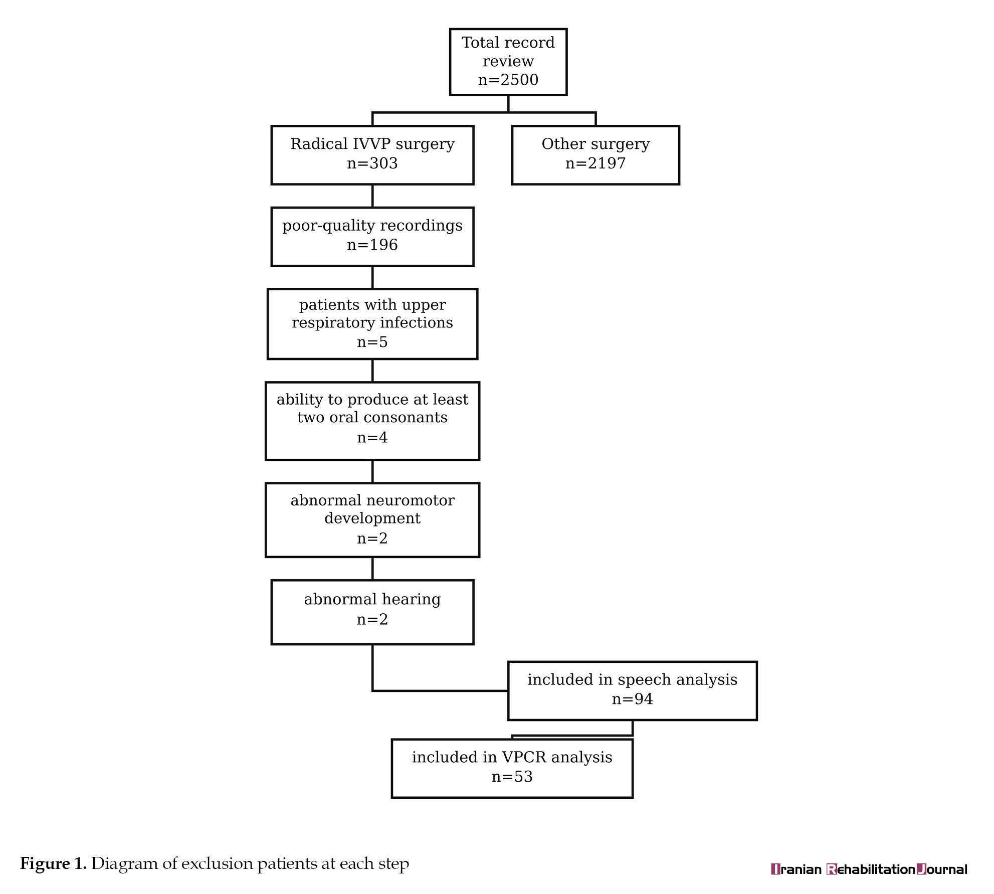
A total of 94 patients who underwent radical IVVP between 2015 and 2020 met the following inclusion criteria: (1) minimum age of 3 years at surgery; (2) at least 6 months between pre- and postoperative assessments; (3) normal hearing at both preoperative and postoperative evaluations; (4) normal neuromotor development (as documented in medical records); and (5) ability to produce at least two oral consonants to allow for accurate assessment of HN independent of the effect of compensatory errors on resonance. Patients with upper respiratory infections or poor-quality recordings were excluded from the analysis. Ultimately, speech characteristics and fistula rate were analyzed for the 94 patients. VPCR analysis was available for 53 patients. Figure 1 illustrates the reasons for exclusion at each step.
 The present study was designed based on a conceptual framework linking 1) anatomical repair by IVVP to 2) physiological VP closure and, ultimately, 3) perceptual resonance outcomes. Within this framework, a partial discordance between objective closure metrics and perceptual speech outcomes was considered an a priori expectation, reflecting the persistence of learned compensatory speech behaviors and multi-structural contributors to resonance despite adequate anatomical repair.
HN was prespecified as the primary outcome measure. All correlations were considered secondary or exploratory analyses.
Data collection
This study was conducted as a single-center retrospective audit of routinely collected clinical data. Pre‑ and post‑operative speech and instrumental assessments were extracted from medical records and analyzed within subjects to evaluate changes following IVVP.
Speech sampling and rating
Pre- and postoperative speech examinations were routinely conducted for every patient at the ICCT. These comprehensive records were made available to the researchers for the audio component of the study. Standardized audio recordings were obtained in a quiet room using Olympus digital media-series recorders, 30 cm from the mouth. Speech tasks included connected speech activities, such as counting and reading a rhyme, as well as structured sentence repetition containing high-pressure consonants, designed in accordance with the universal parameters guidelines [13, 14]. Recordings were anonymized, and two speech-language pathologists performed perceptual evaluation: one with over ten years of experience and the other with three years, using the cleft audit protocol for speech–augmented (CAPS-A) protocol [13]. To ensure blinding, an independent individual not involved in the study randomly coded all recordings.
Ratings were assigned based on CAPS-A criteria
1) HN: 5-point scale (0–4). In this study, to facilitate the interpretation of the results, HN was categorized as ‘no surgery needed’ (0–2), ‘requiring surgery’ (3–4), or ‘improved’ (score reduced from 4 to 3). 2) ANE: 3-point scale (0–2). In this study, to facilitate the interpretation of the results, ANE was categorized as ‘normal’(0) or ‘improved’ (score reduced). 3) NOEs: 3-point scale; categorized as ‘normal’ (0), ‘affected’ (1–2), or ‘improved’ (score reduced). 4) HypoN: 3-point scale (0–2). 5) These variables were compared preoperatively and at least six months postoperatively.
Fistula rate
Presence of oronasal fistula was documented pre- and postoperatively using a binary scale (0=absent, 1=present) based on intraoral examination in the patients’ clinical files.
Velopharyngeal closure ratio
Lateral videofluoroscopy of 53 patients (both pre- and postoperative) was available in the ICCT at the time of the study. The remaining patients were excluded because they lacked either the pre- or postoperative file, or both. Furthermore, the relevant files, particularly for older patients, were not available in the records. Additionally, some patients, especially those with greater functional improvement, were unable or unwilling to undergo postoperative lateral videofluoroscopy, largely because they had to travel from distant areas.
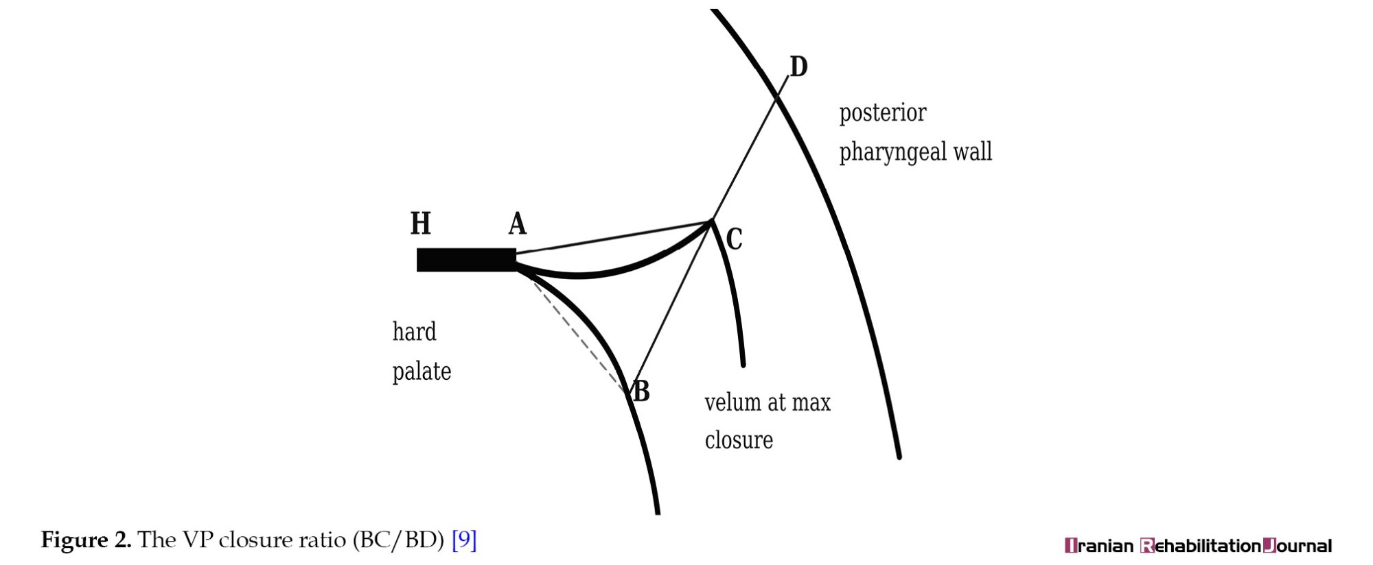
A blinded radiologist calculated VPCR using the ratio of velar movement from rest to closure during /i/ phonation. In this calculation, 1.0 indicates full closure, and 0 indicates no movement [9]. Figure 2 visually represents this measurement.
Data analysis
Data were analyzed using SPSS software, version 24. The Wilcoxon signed-rank test was used to assess pre-post changes in speech parameters, and McNemar’s test was used to assess the fistula rate. The normality of VPCR distribution was assessed using the Shapiro-Wilk test. Consequently, the VPCR data were analyzed using a paired t-test. Inter-rater reliability was calculated using Cohen’s kappa statistic. A Mann-Whitney U test was conducted to compare HN severity distributions between the group with VPCR analysis and the group without VPCR analysis. Ordinal logistic regression was employed to examine predictors of postoperative HN. Statistical significance was set at P<0.05.
Surgical procedure
Four surgeons performed radical IVVP, each with 4 to 7 years of experience with this technique. An operating microscope was employed to facilitate the accurate identification and repositioning of the levator veli palatini muscles, as originally described by Sommerlad et al. [9]. The procedure involved full dissection and meticulous reapproximation of the levator muscle fibers to reestablish a physiological muscle sling orientation.
Results
Patient demographics
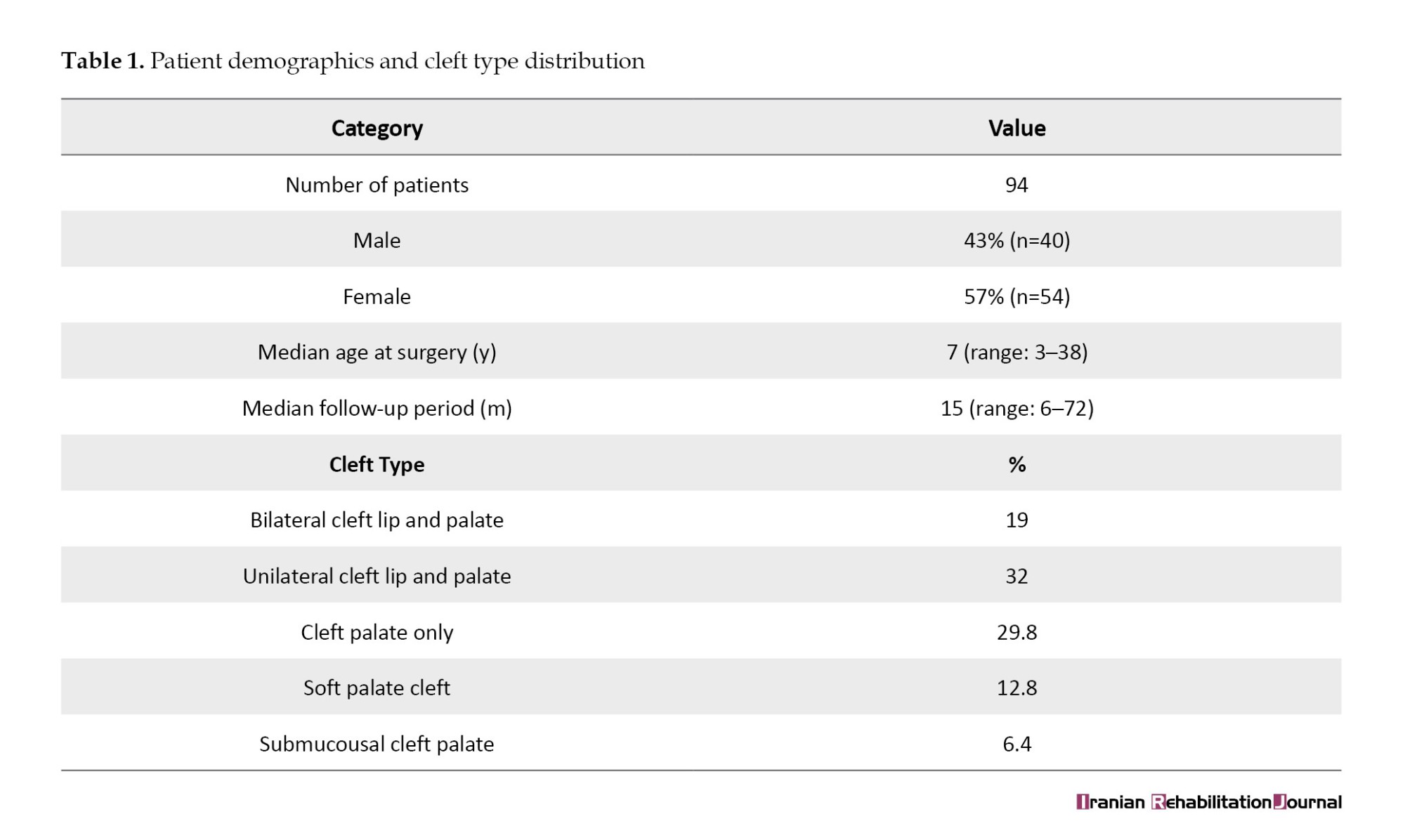
The study included 94 non-syndromic patients (43% male, 57% female). The median age at surgery was 7 years (range: 3–38 years), and the median follow-up was 15 months (range: 6–72). Cleft types included unilateral cleft lip and palate (32%), bilateral cleft lip and palate (19%), complete cleft palate (29.8%), soft palate cleft (12.8%), and submucous cleft (6.4%) (Table 1).
Speech results
Inter-rater agreement was found to be substantial to perfect for all parameters: HN (κ=0.835), HypoN (κ=0.781), ANE (κ=0.92), and NOE (κ=0.84).
The Wilcoxon signed-rank test revealed significant postoperative improvements in HN (P<0.001, r=0.65), ANE (P=0.001, r=0.33), and NOE (P<0.001, r=0.43), while no significant change was observed for HypoN (P=0.603).
Table 2 demonstrates that postoperative outcomes were not dichotomous but distributed along a clinical continuum.
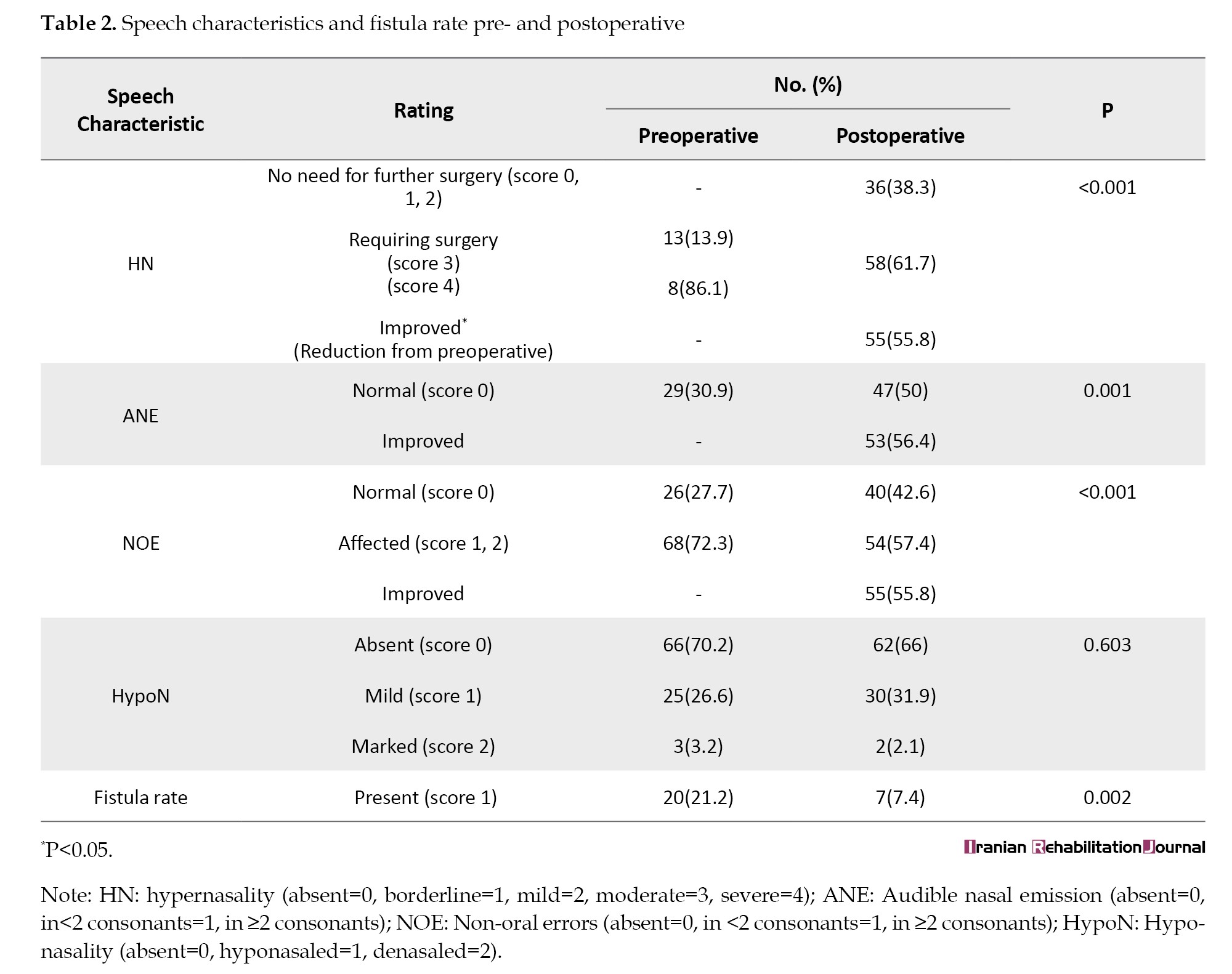
While 55.8% of patients exhibited a reduction in HN severity compared with preoperative levels, this improvement did not uniformly translate into surgical sufficiency. Specifically, 38.3% of patients reached a level of nasality deemed clinically acceptable, obviating the need for further surgical intervention. In contrast, 61.7% of patients continued to demonstrate residual HN of sufficient severity to warrant consideration of additional surgical management. Additionally, 50% of the patients achieved normalized ANE scores.
Fistula rate
Fistula rate decreased significantly from 21.2% (n=20) preoperatively to 7.4% (n=7) postoperatively (P=0.002, [95% CI, 0.07%, 0.52%]), indicating improved structural outcomes.
Velopharyngeal closure ratio
Among 53 patients with pre- and postoperative videofluoroscopy, mean VPCR increased from 0.48±0.28 to 0.70±0.27 (P<0.001, r=0.07). Complete VP closure was achieved in 13 patients. The Shapiro-Wilk test indicated that the data were normally distributed (P>0.05).
The total sample size was divided into two independent groups: Those with VPCR data and those without VPCR data (n=41). A Mann-Whitney U test demonstrated no statistically significant difference in HN between the two groups (U=950.0, P=0.37).
Predictor factors
Ordinal logistic regression was employed to examine the predictors of postoperative HN. The analysis utilized the Logit link function. The test of parallel lines indicated that the proportional odds assumption was satisfied (χ2(15)=13.65, P=0.55).
The model significantly improved prediction of the outcome compared to the intercept-only model (χ2(5)=24.88, P<0.001). The goodness-of-fit test indicated that the model provided an adequate fit to the data, as the deviance statistic was not significant (deviance=121.8, df=191, P=1.00).
These results highlight the significance of preoperative NOE, age at surgery, and postoperative VP closure ratio in predicting postoperative outcomes, while follow-up time does not appear to be a significant factor.
According to Table 3, a significant negative association was observed between preoperative NOE and the outcome.
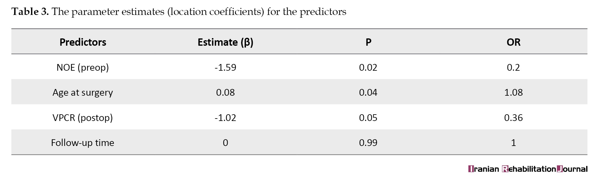
Higher preoperative NOE values were linked to a reduced likelihood of a favorable postoperative outcome. Additionally, age at the time of surgery was positively associated with the outcome.
The postoperative VP closure ratio was also negatively associated with the outcome, suggesting that better postoperative VP closure is associated with a higher probability of a favorable outcome (95% CI, 0.15%, 0.95%).
Follow-up time, however, did not significantly predict the outcomes. The odds ratio (OR) of 1.00 further supports the absence of a relationship between follow-up duration and the outcome.
Discussion
This study evaluated the outcomes of radical IVVP in a relatively large cohort of non-syndromic cleft palate patients. Significant improvements were observed in HN, ANE, and NOEs, along with a substantial reduction in fistula rate and a statistically significant increase in the VPCR. However, a notable proportion of patients continued to demonstrate residual HN or required additional surgical intervention, underscoring the complexity of speech management in cleft palate populations.
The patient cohort’s demographic characteristics, including a mix of cleft types and a median age of seven years at the time of surgery, reflect the heterogeneity often encountered in cleft palate populations. This variability underscores the importance of considering specific anatomical and physiological factors when interpreting the outcomes of secondary surgical interventions.
Speech results
High inter-rater reliability for the perceptual parameters validated the robustness of the rating procedures.
Hypernasality, audible nasal emission, and non-oral errors
Postoperative reductions in HN, ANE, and NOEs confirm that radical IVVP effectively improves VP function in a significant number of patients. Nevertheless, 61.7% of patients still met criteria indicating the need for further surgery, a figure that is higher than those reported in other published studies on palatal re-repair [6, 9]. This discrepancy may be attributed to multiple interacting factors.
1) Patient characteristics and sample composition: Age at the time of surgery is a crucial predictor of outcomes [15-17]. Patients who achieved normal or mild HN underwent surgery at a younger age (mean, 5.7 years) compared to those with persistent severe HN (mean, 15 years). Likewise, patients without postoperative ANE had a lower mean age at surgery. The observed associations highlight the importance of early intervention and the burden of entrenched compensatory errors in older patients.
2) Quality of primary surgeries and case complexity: Studies suggest better outcomes when both primary and secondary surgeries are performed by the same surgeon, particularly when IVVP is used for the primary repair [6, 18, 19]. In this study, many patients had previously undergone one or more suboptimal primary palatoplasties, often outside a multidisciplinary team setting. These “salvage” cases involved extensive scarring or distorted muscle anatomy, complicating re-repair. Rezaei et al. confirmed that many patients in Iran still lack access to interdisciplinary care during early childhood, which can impact subsequent treatment outcomes [20]. Limited surgical records further complicated the evaluation of these cases, and inconsistent primary-surgery quality likely contributed to the lower success rate observed.
3) Surgeon experience and variability: All procedures were performed by four surgeons with 4-7 years’ experience. This contrasts with earlier studies where a single highly experienced surgeon conducted all surgeries [6, 9], which may explain variability in outcomes.
4) Differences in speech scoring systems: In prior studies such as that by Elsherbiny et al., the use of broader scoring systems, such as the Pittsburgh weighted speech score—which includes articulation as well as resonance [6]—may have yielded higher apparent success rates. In contrast, our study used CAPS-A and categorized patients strictly based on resonance and emission outcomes, which may present a more conservative estimate of success. These findings underscore the complexity of evaluating surgical outcomes in VPI and emphasize the multifactorial influences—including age, preoperative speech status, and surgical history—on perceptual speech improvements.
Hyponasality
No significant changes in HypoN were observed postoperatively (P=0.603), which is consistent with the anatomical selectivity of IVVP. Unlike procedures such as pharyngeal flaps or sphincter pharyngoplasties, IVVP avoids altering pharyngeal wall structures, preserving airway patency and reducing the risk of complications, such as obstructive sleep apnea [9]. These findings affirm the relative safety of radical IVVP, particularly in patients at higher risk for airway compromise.
Role of NOEs in predicting postoperative hypernasality
The regression results demonstrated that preoperative NOEs affect residual HN. In other words, speech motor control and articulation patterns play a critical role in determining surgical outcomes. This aligns with studies indicating that NOEs are more than compensatory—they reflect neuromotor or phonological processing challenges that persist even when anatomical closure is restored [21, 22].
Furthermore, NOEs often reflect entrenched compensatory speech habits developed in response to VPI. Even after surgical correction, these patterns may persist unless addressed through targeted speech therapy. Studies suggest that preoperative therapy can reduce compensatory errors and improve postoperative outcomes [23]. Moreover, the severity of NOEs may indicate more severe or long-standing VPI, potentially requiring more complex interventions and yielding lower success rates. Early intervention is therefore critical to prevent entrenched patterns. Future research should explore the impact of combined surgical and therapeutic strategies—especially pre-surgical therapy and neurological factors influencing compensatory articulation—to improve long-term outcomes [24-26].
Age at surgery and postoperative hypernasality
Our findings highlight age as a significant predictor of postoperative speech outcomes. An OR of 1.08 suggests that increasing age is associated with a higher likelihood of more severe postoperative HN. Consistent with this, older patients demonstrated higher rates of HN and ANE. As speech and language patterns solidify with age, the window for optimal surgical impact narrows. Several factors may underlie this association. As children grow older, their speech and language systems become more firmly established. With VP dysfunction, this may reinforce maladaptive articulatory patterns that can persist even after surgical correction. Hence, prolonged exposure to VPI-related deficits before surgical intervention may result in more entrenched speech errors, leading to more severe residual HN postoperatively [15, 16, 27]. Additionally, the findings suggest that IVVP is particularly effective for eliminating VPI in children compared to adults [12]. This emphasizes the importance of early detection and timely surgical referral. Unfortunately, due to referral patterns in Iran, many patients undergo initial surgery in non-specialized centers and present to cleft teams later in childhood, after speech issues have become ingrained.
Lack of detailed surgical records also limited our ability to assess the timing and quality of prior surgeries.
Fistula rate
Our study showed a significant decrease in the fistula rate from 21.2% preoperatively to 7.4% postoperatively (P=0.002), demonstrating the technical reliability of IVVP in providing robust closure. These rates compare favorably with previous studies [28, 29]. However, fistula recurrence in a small subset of patients suggests the need for careful patient selection and further refinement of surgical technique in complex cases. Notably, many of our patients had undergone multiple surgeries before referral, likely contributing to a challenging tissue environment.
Velopharyngeal closure ratio and hypernasality
Although the mean VPCR increased from 0.48 to 0.7 postoperatively and complete closure was achieved in 13 patients, the VPCR and HN results should be interpreted with caution. VPCR demonstrated an inverse association with postoperative HN, suggesting that improved VP closure is linked to reduced HN severity. While the statistical significance was borderline, the observed effect size supports the clinical relevance of VPCR as a potential predictor of speech outcomes. This implies that structural improvement alone does not guarantee perceptual normalization, supporting the view that speech outcomes are multifactorial [24].
Mangal et al. reported a moderately strong negative correlation between the grade of VP port and HN. These results suggest that more severe VPI leads to higher HN, reinforcing the importance of both perceptual and objective assessments in evaluating the impact of VPI on speech [30]. While the systematic review highlights limited and inconsistent evidence concerning the direct association between VP gap size and perceptual nasality, it also underscores the paucity of robust and methodologically rigorous literature addressing the correlation between instrumental findings and clinical speech outcomes [31].
In addition to VPCR, variables such as lateral pharyngeal wall motion, tongue position, oral cavity size, and individual neuromotor control may all affect speech resonance [24, 32, 33]. The mismatch between instrumental and perceptual findings in some cases further underscores the need for combined assessment strategies that go beyond imaging.
Single-view lateral videofluoroscopy is widely used in cleft-related outcome studies to quantify anterior-posterior velar movement and calculate the VPCR. This view provides a reproducible estimate of the relationship between the velum and the posterior pharyngeal wall. However, it does not allow for direct visualization of lateral pharyngeal wall contribution or patterns of complete peripheral closure. In our study, VPCR was measured during sustained /i/ phonation because this vowel consistently elicits maximal and stable velar elevation in a controlled, sustained context, allowing standardized frame selection and measurement reliability [9, 34]. While pressure consonants may better reflect functional speech symptoms of VPI, their dynamic and transient nature makes them less suitable for reproducible radiographic quantification in retrospective image analysis, although VPCR derived from sustained phonation may not fully generalize to connected speech or high-pressure consonant contexts [34, 35].
Follow-up time and severity of hypernasality and audible nasal emission
Follow-up time did not significantly predict speech improvement, echoing the conclusion that early, targeted intervention plays a greater role than follow-up duration alone. Structured, high-quality therapy tailored to the patient’s unique deficits—rather than passive follow-up—is likely to yield better functional outcomes. This finding calls into question protocols that delay postoperative intervention in favor of “wait-and-see” approaches. Although adequate follow‑up is essential to confirm the stability of postoperative speech outcomes, our findings suggest that the severity of HN is primarily determined by factors present at the time of intervention, such as age at surgery, preoperative speech characteristics, and postoperative VP closure. Prolonged follow‑up alone does not appear to further modify these outcomes.
Limitations
The retrospective nature of the study, variability in prior surgeries, the absence of standardized therapy protocols, and the lack of objective tools such as nasometry or nasendoscopy reduce the generalizability and reliability of the results. Furthermore, many patients had incomplete surgical histories due to referrals from external centers, which may introduce bias and reduce data completeness. These factors limit the external validity of the findings and suggest the need for more controlled studies in the future.
Recommendation
To address these limitations, future research should focus on prospective study designs with standardized speech therapy protocols to ensure consistency in treatment. A multi-modal approach to VP assessment, incorporating objective tools such as nasendoscopy and nasometry in addition to VP closure ratio, would provide more accurate and quantifiable data, leading to a better understanding of treatment outcomes. Furthermore, model-based predictors could enhance patient selection and stratification for radical IVVP, improving the overall efficacy of the intervention. Replicating this study with a larger sample size would also increase statistical power, improve generalizability, and provide more robust evidence for the observed findings.
Conclusion
This study supports the effectiveness of IVVP in improving speech outcomes in patients with VPI, although many patients still needed additional surgery to improve speech, especially HN. Significant reductions in HN, ANE, and NOEs were observed postoperatively in a large sample of non-syndromic cleft palate patients. Age at surgery and preoperative articulation patterns were key determinants of postoperative speech improvement, while changes in VP closure should be interpreted alongside perceptual assessments. These findings underscore the need for early intervention, individualized speech therapy, and perceptual assessments as essential components of comprehensive cleft care following secondary surgery.
Ethical Considerations
Compliance with ethical guidelines
This study was approved by the Ethics Committee of Isfahan University of Medical Sciences, Isfahan, Iran (Code: IR.MUI.MED.REC.1395.815).
Funding
The paper was extracted from the master's thesis of Mehri Tavakoli, approved by the Department of Speech Therapy, Faculty of Rehabilitation Sciences, Isfahan University of Medical Sciences, Isfahan, Iran.
Authors' contributions
Software: Mehri Tavakoli and Fatemeh Derakhshandeh, Parisa Rezaei; Formal analysis: Mehri Tavakoli, Fatemeh Derakhshandeh and Parisa Rezaei; Data curation: Mehri Tavakoli, Fatemeh Derakhshandeh, Parisa Rezaei, Hossein Abdali; Supervision: Fatemeh Derakhshandeh, Parisa Rezaei; Project administration: Fatemeh Derakhshandeh; Conceptualization, methodology, resources, validation, visualization, investigation and writing,: All authors.
Conflict of interest
The authors declared no conflict of interest.
Acknowledgments
The authors thank all patients for participating in this study.
References
Velopharyngeal insufficiency (VPI) is characterized by incomplete closure of the velopharyngeal (VP) port during speech or swallowing, typically due to anatomical or functional deficits. Despite advances in primary cleft palate repair techniques, residual VPI persists at a significant rate of 10–40%, frequently leading to speech disorders, such as hypernasality (HN), nasal emission, and compensatory articulation errors [1–5].
Several secondary surgical procedures, including pharyngeal flap, sphincter pharyngoplasty, posterior pharyngeal wall augmentation, and palatal re-repair, have been employed to address persistent VPI [2, 6, 7]. Blacam et al. in a systematic review, reported that after pharyngeal flap surgery, improved resonance and normal resonance were observed in 79.9% and 75.6% of cases, respectively. Similar results were reported for sphincter palatoplasty (78.7% and 61.4%), palatoplasty procedures (73.2% and 57.5%), and pharyngeal wall augmentation (76.9% and 51.3%), respectively [8].
Among these, palatal re-repair has gained attention for its potential to restore velar function while offering lower morbidity and a reduced risk of obstructive sleep apnea. This technique repositions the abnormally oriented levator veli palatini muscles to a more functional posterior alignment and was initially developed and validated in a large series of palatal re-repairs, specifically targeting restoration of VP function. The technique achieves this through radical/extensive dissection to meticulously isolate and reattach the individual levator muscles directly to each other, effectively reconstructing the velar muscular sling. This anatomical control and direct levator-to-levator repair are key factors contributing to its proven efficacy in challenging secondary cases [6-11].
Palatal re‑repair is not a novel surgical technique but represents an evolution of intravelar veloplasty (IVVP) principles described for several decades. Historically, this approach has been reported under different terminologies, including radical IVVP and secondary muscle repositioning, rather than as a distinct category. Radical IVVP, a variant of palatal re‑repair, focuses on complete dissection and anatomical repositioning of the levator veli palatini muscles to restore a functional velar sling.
Sommerlad et al. reported that over 80% of patients with VPI exhibited either normal nasality and nasal airflow or only mild, inconsistent HN and nasal emission (NE) following palatal re-repair [9]. Elsherbiny et al. reported highly significant improvements in nasality, NE, articulation, and overall speech scores following palatal re-repair. Postoperatively, 66.7% of patients achieved normal or borderline speech, whereas 24.3% exhibited further improvement [6]. In another study, 65% of patients achieved surgical success characterized by the elimination or reduction of HN; 55% demonstrated adequate nasality, while 10% showed a decrease in HN from moderate to mild. Postoperative normal nasality was observed only among children (73%), indicating superior outcomes in younger patients [12]. A systematic review and meta-analysis assessing the effectiveness of palatal re-repair for VPI treatment found that 61% of patients achieved a resolution of consistent HN following the procedure. The technique of palatal re-repair encompasses three main categories: Furlow double-opposing Z-plasty, radical IVVP, and radical IVVP with palatal lengthening [7]. This paper specifically focuses on the outcomes associated with radical IVVP.
Radical IVVP, a variant of palatal re-repair, has been performed by the Isfahan craniofacial and cleft research center (ICCT) since 2010 to address anatomical abnormalities of the palate and enhance speech and resonance outcomes. Given that perceptual evaluation is considered the gold standard for assessing speech and resonance features [1], it is crucial to evaluate the effectiveness of the radical IVVP technique offered by the ICCT through perceptual methods. In addition to perceptual assessments, instrumental evaluations, such as videofluoroscopy, provide valuable insights into the structure and function of the VP mechanism [1]. Despite varying results reported by different researchers regarding the outcome measures of this technique in correcting speech symptoms, no comprehensive audit study has been conducted to evaluate its effectiveness in Iran, specifically at ICCT. Given the continuous advancements and modifications in surgical techniques over the years, it is vital to assess the current outcomes and provide essential feedback to the surgical team for quality improvement.
This study retrospectively evaluated the outcomes of radical IVVP in non-syndromic cleft palate patients. The primary objectives included assessing perceptual speech parameters, such as HN, audible nasal emission (ANE), hyponasality (HypoN), and non-oral errors (NOEs). A secondary objective was to examine the NOEs, age at surgery, VP closure ratio (VPCR), and follow-up time as predictor factors for postoperative HN following radical IVVP.
Materials and Methods
Study design and participants
Medical records of 2,500 cleft palate patients with VPI were reviewed. Of these, 303 patients underwent radical IVVP as the final secondary surgical procedure performed by the team’s surgeons between 2015 and 2020.
A total of 94 patients who underwent radical IVVP between 2015 and 2020 met the following inclusion criteria: (1) minimum age of 3 years at surgery; (2) at least 6 months between pre- and postoperative assessments; (3) normal hearing at both preoperative and postoperative evaluations; (4) normal neuromotor development (as documented in medical records); and (5) ability to produce at least two oral consonants to allow for accurate assessment of HN independent of the effect of compensatory errors on resonance. Patients with upper respiratory infections or poor-quality recordings were excluded from the analysis. Ultimately, speech characteristics and fistula rate were analyzed for the 94 patients. VPCR analysis was available for 53 patients. Figure 1 illustrates the reasons for exclusion at each step.
HN was prespecified as the primary outcome measure. All correlations were considered secondary or exploratory analyses.
Data collection
This study was conducted as a single-center retrospective audit of routinely collected clinical data. Pre‑ and post‑operative speech and instrumental assessments were extracted from medical records and analyzed within subjects to evaluate changes following IVVP.
Speech sampling and rating
Pre- and postoperative speech examinations were routinely conducted for every patient at the ICCT. These comprehensive records were made available to the researchers for the audio component of the study. Standardized audio recordings were obtained in a quiet room using Olympus digital media-series recorders, 30 cm from the mouth. Speech tasks included connected speech activities, such as counting and reading a rhyme, as well as structured sentence repetition containing high-pressure consonants, designed in accordance with the universal parameters guidelines [13, 14]. Recordings were anonymized, and two speech-language pathologists performed perceptual evaluation: one with over ten years of experience and the other with three years, using the cleft audit protocol for speech–augmented (CAPS-A) protocol [13]. To ensure blinding, an independent individual not involved in the study randomly coded all recordings.
Ratings were assigned based on CAPS-A criteria
1) HN: 5-point scale (0–4). In this study, to facilitate the interpretation of the results, HN was categorized as ‘no surgery needed’ (0–2), ‘requiring surgery’ (3–4), or ‘improved’ (score reduced from 4 to 3). 2) ANE: 3-point scale (0–2). In this study, to facilitate the interpretation of the results, ANE was categorized as ‘normal’(0) or ‘improved’ (score reduced). 3) NOEs: 3-point scale; categorized as ‘normal’ (0), ‘affected’ (1–2), or ‘improved’ (score reduced). 4) HypoN: 3-point scale (0–2). 5) These variables were compared preoperatively and at least six months postoperatively.
Fistula rate
Presence of oronasal fistula was documented pre- and postoperatively using a binary scale (0=absent, 1=present) based on intraoral examination in the patients’ clinical files.
Velopharyngeal closure ratio
Lateral videofluoroscopy of 53 patients (both pre- and postoperative) was available in the ICCT at the time of the study. The remaining patients were excluded because they lacked either the pre- or postoperative file, or both. Furthermore, the relevant files, particularly for older patients, were not available in the records. Additionally, some patients, especially those with greater functional improvement, were unable or unwilling to undergo postoperative lateral videofluoroscopy, largely because they had to travel from distant areas.
A blinded radiologist calculated VPCR using the ratio of velar movement from rest to closure during /i/ phonation. In this calculation, 1.0 indicates full closure, and 0 indicates no movement [9]. Figure 2 visually represents this measurement.
Data analysis
Data were analyzed using SPSS software, version 24. The Wilcoxon signed-rank test was used to assess pre-post changes in speech parameters, and McNemar’s test was used to assess the fistula rate. The normality of VPCR distribution was assessed using the Shapiro-Wilk test. Consequently, the VPCR data were analyzed using a paired t-test. Inter-rater reliability was calculated using Cohen’s kappa statistic. A Mann-Whitney U test was conducted to compare HN severity distributions between the group with VPCR analysis and the group without VPCR analysis. Ordinal logistic regression was employed to examine predictors of postoperative HN. Statistical significance was set at P<0.05.
Surgical procedure
Four surgeons performed radical IVVP, each with 4 to 7 years of experience with this technique. An operating microscope was employed to facilitate the accurate identification and repositioning of the levator veli palatini muscles, as originally described by Sommerlad et al. [9]. The procedure involved full dissection and meticulous reapproximation of the levator muscle fibers to reestablish a physiological muscle sling orientation.
Results
Patient demographics
The study included 94 non-syndromic patients (43% male, 57% female). The median age at surgery was 7 years (range: 3–38 years), and the median follow-up was 15 months (range: 6–72). Cleft types included unilateral cleft lip and palate (32%), bilateral cleft lip and palate (19%), complete cleft palate (29.8%), soft palate cleft (12.8%), and submucous cleft (6.4%) (Table 1).
Speech results
Inter-rater agreement was found to be substantial to perfect for all parameters: HN (κ=0.835), HypoN (κ=0.781), ANE (κ=0.92), and NOE (κ=0.84).
The Wilcoxon signed-rank test revealed significant postoperative improvements in HN (P<0.001, r=0.65), ANE (P=0.001, r=0.33), and NOE (P<0.001, r=0.43), while no significant change was observed for HypoN (P=0.603).
Table 2 demonstrates that postoperative outcomes were not dichotomous but distributed along a clinical continuum.
While 55.8% of patients exhibited a reduction in HN severity compared with preoperative levels, this improvement did not uniformly translate into surgical sufficiency. Specifically, 38.3% of patients reached a level of nasality deemed clinically acceptable, obviating the need for further surgical intervention. In contrast, 61.7% of patients continued to demonstrate residual HN of sufficient severity to warrant consideration of additional surgical management. Additionally, 50% of the patients achieved normalized ANE scores.
Fistula rate
Fistula rate decreased significantly from 21.2% (n=20) preoperatively to 7.4% (n=7) postoperatively (P=0.002, [95% CI, 0.07%, 0.52%]), indicating improved structural outcomes.
Velopharyngeal closure ratio
Among 53 patients with pre- and postoperative videofluoroscopy, mean VPCR increased from 0.48±0.28 to 0.70±0.27 (P<0.001, r=0.07). Complete VP closure was achieved in 13 patients. The Shapiro-Wilk test indicated that the data were normally distributed (P>0.05).
The total sample size was divided into two independent groups: Those with VPCR data and those without VPCR data (n=41). A Mann-Whitney U test demonstrated no statistically significant difference in HN between the two groups (U=950.0, P=0.37).
Predictor factors
Ordinal logistic regression was employed to examine the predictors of postoperative HN. The analysis utilized the Logit link function. The test of parallel lines indicated that the proportional odds assumption was satisfied (χ2(15)=13.65, P=0.55).
The model significantly improved prediction of the outcome compared to the intercept-only model (χ2(5)=24.88, P<0.001). The goodness-of-fit test indicated that the model provided an adequate fit to the data, as the deviance statistic was not significant (deviance=121.8, df=191, P=1.00).
These results highlight the significance of preoperative NOE, age at surgery, and postoperative VP closure ratio in predicting postoperative outcomes, while follow-up time does not appear to be a significant factor.
According to Table 3, a significant negative association was observed between preoperative NOE and the outcome.
Higher preoperative NOE values were linked to a reduced likelihood of a favorable postoperative outcome. Additionally, age at the time of surgery was positively associated with the outcome.
The postoperative VP closure ratio was also negatively associated with the outcome, suggesting that better postoperative VP closure is associated with a higher probability of a favorable outcome (95% CI, 0.15%, 0.95%).
Follow-up time, however, did not significantly predict the outcomes. The odds ratio (OR) of 1.00 further supports the absence of a relationship between follow-up duration and the outcome.
Discussion
This study evaluated the outcomes of radical IVVP in a relatively large cohort of non-syndromic cleft palate patients. Significant improvements were observed in HN, ANE, and NOEs, along with a substantial reduction in fistula rate and a statistically significant increase in the VPCR. However, a notable proportion of patients continued to demonstrate residual HN or required additional surgical intervention, underscoring the complexity of speech management in cleft palate populations.
The patient cohort’s demographic characteristics, including a mix of cleft types and a median age of seven years at the time of surgery, reflect the heterogeneity often encountered in cleft palate populations. This variability underscores the importance of considering specific anatomical and physiological factors when interpreting the outcomes of secondary surgical interventions.
Speech results
High inter-rater reliability for the perceptual parameters validated the robustness of the rating procedures.
Hypernasality, audible nasal emission, and non-oral errors
Postoperative reductions in HN, ANE, and NOEs confirm that radical IVVP effectively improves VP function in a significant number of patients. Nevertheless, 61.7% of patients still met criteria indicating the need for further surgery, a figure that is higher than those reported in other published studies on palatal re-repair [6, 9]. This discrepancy may be attributed to multiple interacting factors.
1) Patient characteristics and sample composition: Age at the time of surgery is a crucial predictor of outcomes [15-17]. Patients who achieved normal or mild HN underwent surgery at a younger age (mean, 5.7 years) compared to those with persistent severe HN (mean, 15 years). Likewise, patients without postoperative ANE had a lower mean age at surgery. The observed associations highlight the importance of early intervention and the burden of entrenched compensatory errors in older patients.
2) Quality of primary surgeries and case complexity: Studies suggest better outcomes when both primary and secondary surgeries are performed by the same surgeon, particularly when IVVP is used for the primary repair [6, 18, 19]. In this study, many patients had previously undergone one or more suboptimal primary palatoplasties, often outside a multidisciplinary team setting. These “salvage” cases involved extensive scarring or distorted muscle anatomy, complicating re-repair. Rezaei et al. confirmed that many patients in Iran still lack access to interdisciplinary care during early childhood, which can impact subsequent treatment outcomes [20]. Limited surgical records further complicated the evaluation of these cases, and inconsistent primary-surgery quality likely contributed to the lower success rate observed.
3) Surgeon experience and variability: All procedures were performed by four surgeons with 4-7 years’ experience. This contrasts with earlier studies where a single highly experienced surgeon conducted all surgeries [6, 9], which may explain variability in outcomes.
4) Differences in speech scoring systems: In prior studies such as that by Elsherbiny et al., the use of broader scoring systems, such as the Pittsburgh weighted speech score—which includes articulation as well as resonance [6]—may have yielded higher apparent success rates. In contrast, our study used CAPS-A and categorized patients strictly based on resonance and emission outcomes, which may present a more conservative estimate of success. These findings underscore the complexity of evaluating surgical outcomes in VPI and emphasize the multifactorial influences—including age, preoperative speech status, and surgical history—on perceptual speech improvements.
Hyponasality
No significant changes in HypoN were observed postoperatively (P=0.603), which is consistent with the anatomical selectivity of IVVP. Unlike procedures such as pharyngeal flaps or sphincter pharyngoplasties, IVVP avoids altering pharyngeal wall structures, preserving airway patency and reducing the risk of complications, such as obstructive sleep apnea [9]. These findings affirm the relative safety of radical IVVP, particularly in patients at higher risk for airway compromise.
Role of NOEs in predicting postoperative hypernasality
The regression results demonstrated that preoperative NOEs affect residual HN. In other words, speech motor control and articulation patterns play a critical role in determining surgical outcomes. This aligns with studies indicating that NOEs are more than compensatory—they reflect neuromotor or phonological processing challenges that persist even when anatomical closure is restored [21, 22].
Furthermore, NOEs often reflect entrenched compensatory speech habits developed in response to VPI. Even after surgical correction, these patterns may persist unless addressed through targeted speech therapy. Studies suggest that preoperative therapy can reduce compensatory errors and improve postoperative outcomes [23]. Moreover, the severity of NOEs may indicate more severe or long-standing VPI, potentially requiring more complex interventions and yielding lower success rates. Early intervention is therefore critical to prevent entrenched patterns. Future research should explore the impact of combined surgical and therapeutic strategies—especially pre-surgical therapy and neurological factors influencing compensatory articulation—to improve long-term outcomes [24-26].
Age at surgery and postoperative hypernasality
Our findings highlight age as a significant predictor of postoperative speech outcomes. An OR of 1.08 suggests that increasing age is associated with a higher likelihood of more severe postoperative HN. Consistent with this, older patients demonstrated higher rates of HN and ANE. As speech and language patterns solidify with age, the window for optimal surgical impact narrows. Several factors may underlie this association. As children grow older, their speech and language systems become more firmly established. With VP dysfunction, this may reinforce maladaptive articulatory patterns that can persist even after surgical correction. Hence, prolonged exposure to VPI-related deficits before surgical intervention may result in more entrenched speech errors, leading to more severe residual HN postoperatively [15, 16, 27]. Additionally, the findings suggest that IVVP is particularly effective for eliminating VPI in children compared to adults [12]. This emphasizes the importance of early detection and timely surgical referral. Unfortunately, due to referral patterns in Iran, many patients undergo initial surgery in non-specialized centers and present to cleft teams later in childhood, after speech issues have become ingrained.
Lack of detailed surgical records also limited our ability to assess the timing and quality of prior surgeries.
Fistula rate
Our study showed a significant decrease in the fistula rate from 21.2% preoperatively to 7.4% postoperatively (P=0.002), demonstrating the technical reliability of IVVP in providing robust closure. These rates compare favorably with previous studies [28, 29]. However, fistula recurrence in a small subset of patients suggests the need for careful patient selection and further refinement of surgical technique in complex cases. Notably, many of our patients had undergone multiple surgeries before referral, likely contributing to a challenging tissue environment.
Velopharyngeal closure ratio and hypernasality
Although the mean VPCR increased from 0.48 to 0.7 postoperatively and complete closure was achieved in 13 patients, the VPCR and HN results should be interpreted with caution. VPCR demonstrated an inverse association with postoperative HN, suggesting that improved VP closure is linked to reduced HN severity. While the statistical significance was borderline, the observed effect size supports the clinical relevance of VPCR as a potential predictor of speech outcomes. This implies that structural improvement alone does not guarantee perceptual normalization, supporting the view that speech outcomes are multifactorial [24].
Mangal et al. reported a moderately strong negative correlation between the grade of VP port and HN. These results suggest that more severe VPI leads to higher HN, reinforcing the importance of both perceptual and objective assessments in evaluating the impact of VPI on speech [30]. While the systematic review highlights limited and inconsistent evidence concerning the direct association between VP gap size and perceptual nasality, it also underscores the paucity of robust and methodologically rigorous literature addressing the correlation between instrumental findings and clinical speech outcomes [31].
In addition to VPCR, variables such as lateral pharyngeal wall motion, tongue position, oral cavity size, and individual neuromotor control may all affect speech resonance [24, 32, 33]. The mismatch between instrumental and perceptual findings in some cases further underscores the need for combined assessment strategies that go beyond imaging.
Single-view lateral videofluoroscopy is widely used in cleft-related outcome studies to quantify anterior-posterior velar movement and calculate the VPCR. This view provides a reproducible estimate of the relationship between the velum and the posterior pharyngeal wall. However, it does not allow for direct visualization of lateral pharyngeal wall contribution or patterns of complete peripheral closure. In our study, VPCR was measured during sustained /i/ phonation because this vowel consistently elicits maximal and stable velar elevation in a controlled, sustained context, allowing standardized frame selection and measurement reliability [9, 34]. While pressure consonants may better reflect functional speech symptoms of VPI, their dynamic and transient nature makes them less suitable for reproducible radiographic quantification in retrospective image analysis, although VPCR derived from sustained phonation may not fully generalize to connected speech or high-pressure consonant contexts [34, 35].
Follow-up time and severity of hypernasality and audible nasal emission
Follow-up time did not significantly predict speech improvement, echoing the conclusion that early, targeted intervention plays a greater role than follow-up duration alone. Structured, high-quality therapy tailored to the patient’s unique deficits—rather than passive follow-up—is likely to yield better functional outcomes. This finding calls into question protocols that delay postoperative intervention in favor of “wait-and-see” approaches. Although adequate follow‑up is essential to confirm the stability of postoperative speech outcomes, our findings suggest that the severity of HN is primarily determined by factors present at the time of intervention, such as age at surgery, preoperative speech characteristics, and postoperative VP closure. Prolonged follow‑up alone does not appear to further modify these outcomes.
Limitations
The retrospective nature of the study, variability in prior surgeries, the absence of standardized therapy protocols, and the lack of objective tools such as nasometry or nasendoscopy reduce the generalizability and reliability of the results. Furthermore, many patients had incomplete surgical histories due to referrals from external centers, which may introduce bias and reduce data completeness. These factors limit the external validity of the findings and suggest the need for more controlled studies in the future.
Recommendation
To address these limitations, future research should focus on prospective study designs with standardized speech therapy protocols to ensure consistency in treatment. A multi-modal approach to VP assessment, incorporating objective tools such as nasendoscopy and nasometry in addition to VP closure ratio, would provide more accurate and quantifiable data, leading to a better understanding of treatment outcomes. Furthermore, model-based predictors could enhance patient selection and stratification for radical IVVP, improving the overall efficacy of the intervention. Replicating this study with a larger sample size would also increase statistical power, improve generalizability, and provide more robust evidence for the observed findings.
Conclusion
This study supports the effectiveness of IVVP in improving speech outcomes in patients with VPI, although many patients still needed additional surgery to improve speech, especially HN. Significant reductions in HN, ANE, and NOEs were observed postoperatively in a large sample of non-syndromic cleft palate patients. Age at surgery and preoperative articulation patterns were key determinants of postoperative speech improvement, while changes in VP closure should be interpreted alongside perceptual assessments. These findings underscore the need for early intervention, individualized speech therapy, and perceptual assessments as essential components of comprehensive cleft care following secondary surgery.
Ethical Considerations
Compliance with ethical guidelines
This study was approved by the Ethics Committee of Isfahan University of Medical Sciences, Isfahan, Iran (Code: IR.MUI.MED.REC.1395.815).
Funding
The paper was extracted from the master's thesis of Mehri Tavakoli, approved by the Department of Speech Therapy, Faculty of Rehabilitation Sciences, Isfahan University of Medical Sciences, Isfahan, Iran.
Authors' contributions
Software: Mehri Tavakoli and Fatemeh Derakhshandeh, Parisa Rezaei; Formal analysis: Mehri Tavakoli, Fatemeh Derakhshandeh and Parisa Rezaei; Data curation: Mehri Tavakoli, Fatemeh Derakhshandeh, Parisa Rezaei, Hossein Abdali; Supervision: Fatemeh Derakhshandeh, Parisa Rezaei; Project administration: Fatemeh Derakhshandeh; Conceptualization, methodology, resources, validation, visualization, investigation and writing,: All authors.
Conflict of interest
The authors declared no conflict of interest.
Acknowledgments
The authors thank all patients for participating in this study.
References
- Kummer AW, Kummer AW. Cleft palate and craniofacial anomalies: effects on speech and resonance. Clifton Park: Thomson Delmar Learning; 2008. [Link]
- Naran S, Miller M, Shakir S, Ware B, Camison L, Ford M, et al. Nonsyndromic craniosynostosis and associated abnormal speech and language development. Plastic and Reconstructive Surgery. 2017; 140(1):62e-9. [DOI:10.1097/PRS.0000000000003423] [PMID]
- Mapar D, Khanlar F, Sadeghi S, Abdali H, Memarzadeh M, Davari HA, et al. The incidence of velopharyngeal insufficiency and oronasal fistula after primary palatal surgery with Sommerlad intravelar veloplasty: A retrospective study in Isfahan Cleft Care Team. International Journal of Pediatric Otorhinolaryngology. 2019; 120:6-10. [DOI:10.1016/j.ijporl.2018.12.035] [PMID]
- Ahti V, Alaluusua S, Vuola P, Rautio J, Leikola J, Saarikko A. Palatal re-repair with Z-plasty in treatment of velopharyngeal insufficiency of syndromic and nonsyndromic patients with cleft palate. Journal of Craniofacial Surgery. 2021; 32(2):685-90. [DOI:10.1097/SCS.0000000000007343] [PMID]
- Reddy SR, Subramaniyan B, Nagarajan R. Studying the impact of cleft of lip and palate among adults using the International classification of functioning, disability and health framework. Journal of Cleft Lip Palate and Craniofacial Anomalies. 2017; 4(2):125-37. [DOI:10.4103/jclpca.jclpca_12_17]
- Elsherbiny A, Amerson M, Sconyers L, Grant JH. Outcome of palate re-repair with radical repositioning of the levator muscle sling as a first-line strategy in postpalatoplasty velopharyngeal incompetence management protocol. Plastic & Reconstructive Surgery. 2018; 141(4):984-91. [DOI:10.1097/PRS.0000000000004236] [PMID]
- Kurnik NM, Weidler EM, Lien KM, Cordero KN, Williams JL, Temkit MH, et al. The effectiveness of palate re-repair for treating velopharyngeal insufficiency: A systematic review and meta-analysis. The Cleft Palate Craniofacial Journal. 2020; 57(7):860-71. [DOI:10.1177/1055665620902883] [PMID] [PMCID]
- de Blacam C, Smith S, Orr D. Surgery for velopharyngeal dysfunction: A systematic review of interventions and outcomes. The Cleft Palate-Craniofacial Journal. 2018; 55(3):405-22.[DOI:10.1177/1055665617735102] [PMID]
- Sommerlad BC, Henley M, Birch M, Harland K, Moiemen N, Boorman J. Cleft palate re-repair—a clinical and radiographic study of 32 consecutive cases. British Journal of Plastic Surgery. 1994; 47(6):406-10. [DOI:10.1016/0007-1226(94)90068-X] [PMID]
- Mehendale FV, Lane R, Laverty A, Dinwiddie R, Sommerlad BC. Effect of palate re-repairs and hynes pharyngoplasties on pediatric airways: An analysis of preoperative and postoperative cardiorespiratory sleep studies. The Cleft Palate-Craniofacial Journal. 2013; 50(3):257-67. [DOI:10.1597/11-198] [PMID]
- Grant III JH, Braswell AC, Soto E. Pros and cons of the Sommerlad and Furlow palate repair techniques. Frontiers of Oral and Maxillofacial Medicine. 2024; 6:14. [DOI:10.21037/fomm-22-72]
- Araújo BM, da Silva AS, Bertier CE, de Sousa Brosco TV, Yamashita RP, Sampaio-Teixeira AC, et al. Palate re-repair for velopharyngeal insufficiency treatment: A long-term auditory-perceptual assessment of speech. Perspectives of the ASHA Special Interest Groups. 2023; 8(5):959-68. [DOI:10.1044/2022_PERSP-22-00079]
- John A, Sell D, Sweeney T, Harding-Bell A, Williams A. The cleft audit protocol for speech—augmented: A validated and reliable measure for auditing cleft speech. The Cleft Palate-Craniofacial Journal. 2006; 43(3):272-88. [DOI:10.1597/04-141.1] [PMID]
- Amirian A, Derakhshandeh F, Salehi A, Soleimani B. Evaluating Intra-and inter-rater reliability for. Journal of Research in Rehabilitation Sciences. 2011; 7(4):470-6. [Link]
- Peterson-Falzone SJ, Hardin-Jones MA, Karnell MP. Cleft Palate Speech. St. Louis: Mosby; 2010. [Link]
- Sell D. Issues in perceptual speech analysis in cleft palate and related disorders: A review. International Journal of Language & Communication Disorders. 2005; 40(2):103-21. [DOI:10.1080/13682820400016522] [PMID]
- Cheng X, Bo Z, Yin H, Yang K, Li J, Shi B. Age and preoperative velar closure ratio are significantly associated with surgical outcome of Furlow double-opposing Z-plasty in palatal re-repair. Journal of Oral and Maxillofacial Surgery. 2020; 78(3):431-9. [DOI:10.1016/j.joms.2019.09.023] [PMID]
- Elsherbiny A, Gelany A, Mazeed AS, Mostafa E, Ahmed MA, Allam KA, et al. Buccinator re-repair (Bs+ Re: IVVP): a combined procedure to maximize the palate form and function in difficult VPI cases. The Cleft Palate-Craniofacial Journal. 2020; 57(5):543-51. [DOI:10.1177/1055665619900621] [PMID]
- Varghese D, Datta S, Varghese A. Use of buccal myomucosal flap for palatal lengthening in cleft palate patient: Experience of 20 cases. Contemporary Clinical Dentistry. 2015; 6(Suppl 1):S36. [DOI:10.4103/0976-237X.152935] [PMID] [PMCID]
- Rezaei P, Poorjavad M, Abdali H. Speech outcomes after palatal closure in 3–7-year-old children. Brazilian Journal of Otorhinolaryngology. 2022; 88(4):594-601. [DOI:10.1016/j.bjorl.2020.08.005] [PMID] [PMCID]
- Dubey AK, Prasanna SM, Dandapat S. Spectral analysis based objective assessment of hypernasality in cleft palate speech: A review. Circuits, Systems, and Signal Processing. 2025; 44(6):4133-66. [DOI:10.1007/s00034-025-03007-x]
- Boyce JO, Kilpatrick N, Morgan AT. Speech and language characteristics in individuals with nonsyndromic submucous cleft palate—A systematic review. Child: Care, Health and Development. 2018; 44(6):818-31. [DOI:10.1111/cch.12613] [PMID]
- Ghandour H, Kaddah F, Abdelhamid A, Elbarbary A. Effects of preoperative speech therapy on the speech intelligibility of patients with velopharyngeal incompetence. Egyptian Journal of Otolaryngology. 2013; 29(4):269-76. [Link]
- Kuehn DP, Moller KT. Speech and language issues in the cleft palate population: The state of the art. The Cleft Palate-Craniofacial Journal. 2000; 37(4):1-35. [DOI:10.1597/1545-1569_2000_037_0348_saliit_2.3.co_2]
- Derakhshandeh F, Nikmaram M, Hosseinabad HH, Memarzadeh M, Taheri M, Omrani M, et al. Speech characteristics after articulation therapy in children with cleft palate and velopharyngeal dysfunction–A single case experimental design. International Journal of Pediatric Otorhinolaryngology. 2016; 86:104-13. [DOI:10.1016/j.ijporl.2016.04.025] [PMID]
- Hosseinabad HH, Xing Y, Kemp M. A retrospective analysis of factors affecting speech production in school-aged children with cleft palate (+-cleft lip). International Journal of Pediatric Otorhinolaryngology. 2024; 182:112029.[DOI:10.1016/j.ijporl.2024.112029] [PMID]
- Baek RM, Kim BK, Jeong JH, Ahn T, Park M, Han J. The effect of age at surgery and compensatory articulation on speech outcome in submucous cleft palate patients treated with double-opposing Z-plasty: A 10-year experience. Journal of Plastic, Reconstructive & Aesthetic Surgery. 2017; 70(5):646-52. [DOI:10.1016/j.bjps.2016.12.025] [PMID]
- Chandra Sekar, PK, Veerabathiran R. Current advances in surgical techniques for secondary cleft palate repair: A systematic review. The Cleft Palate Craniofacial Journal; 2025; 10556656251409261. [DOI:10.1177/10556656251409261] [PMID]
- Baillie L, Sell D. Benchmarking speech, velopharyngeal function outcomes and surgical characteristics following the Sommerlad protocol and palate repair technique. The Cleft Palate Craniofacial Journal. 2020; 57(10):1197-215.[DOI:10.1177/1055665620923925] [PMID]
- Mangal M, Kumar P, Munjal S, Sharma RK. Endoscopic evaluation of velopharyngeal function in cleft lip palate patients–A correlation with speech analysis. Journal of Plastic, Reconstructive & Aesthetic Surgery. 2023; 77:170-6.[DOI:10.1016/j.bjps.2022.10.041] [PMID]
- Paniagua LM, Signorini AV, da Costa SS, Collares MV, Dornelles S. Velopharyngeal dysfunction: A systematic review of major instrumental and auditory-perceptual assessments. International Archives of Otorhinolaryngology. 2013; 17(03):251-6. [DOI:10.7162/S1809-97772013000300004] [PMID] [PMCID]
- Grabski K, Lamalle L, Vilain C, Schwartz J, Vallée N, Tropres I, et al. Functional MRI assessment of orofacial articulators: neural correlates of lip, jaw, larynx, and tongue movements. Human Brain Mapping; 2012: 33(10):2306-21. [DOI:10.1002/hbm.21363] [PMID]
- Nagarajan R, Savitha VH, Subramaniyan B. Communication disorders in individuals with cleft lip and palate: An overview. Indian Journal of Plastic Surgery. 2009; 42(S 01):S137-43. [DOI:10.4103/0970-0358.57199] [PMID]
- Sullivan SR, Taylor HO, Stiles E, Nuss RC, Balkin DM, Mulliken JB. Videofluoroscopy versus nasopharyngoscopy of lateral pharyngeal wall movement for assessment of velopharyngeal insufficiency. Journal of Craniofacial Surgery. 2023; 34(1):40-4. [DOI:10.1097/SCS.0000000000008945] [PMID]
- Eshghi M, Vallino LD, Baylis AL, Preisser JS, Zajac DJ. Velopharyngeal status of stop consonants and vowels produced by young children with and without repaired cleft palate at 12, 14, and 18 months of age: A preliminary analysis. Journal of Speech, Language, and Hearing Research. 2017; 60(6):1467-76. [DOI:10.1044/2016_JSLHR-S-16-0259] [PMID]
Article type: Original Research Articles |
Subject:
Speech therapy
Received: 2025/12/16 | Accepted: 2026/05/2 | Published: 2026/09/26
Received: 2025/12/16 | Accepted: 2026/05/2 | Published: 2026/09/26
Send email to the article author

Copyright © The Author(s);
This is an open access article distributed under the terms of the Creative Commons Attribution-NonCommercial 4.0 (CC-By-NC 4.0), which permits use, distribution, and reproduction in any medium, provided the original work is properly cited and is not used for commercial purposes.
Contact Information
