
988368
Sat, Jun 27, 2026

Volume 24, Issue 1 (March 2026)
Iranian Rehabilitation Journal 2026, 24(1): 27-50 |
Back to browse issues page


Download citation:
BibTeX | RIS | EndNote | Medlars | ProCite | Reference Manager | RefWorks
Send citation to:


BibTeX | RIS | EndNote | Medlars | ProCite | Reference Manager | RefWorks
Send citation to:
Salsali M, Sayyadi P, Rajabi E, Sheikhhoseini R, Ebrahimi E, Guess T M. Fatigue-induced Changes in Lower Extremity Landing Kinematics: A Systematic Review and Meta-analysis. Iranian Rehabilitation Journal 2026; 24 (1) :27-50
URL: http://irj.uswr.ac.ir/article-1-2626-en.html
URL: http://irj.uswr.ac.ir/article-1-2626-en.html
Fatigue-induced Changes in Lower Extremity Landing Kinematics: A Systematic Review and Meta-analysis
Mohammad Salsali1 

 , Parisa Sayyadi2 , Elnaz Rajabi3 , Rahman Sheikhhoseini *3 , Ebrahim Ebrahimi2 , Trent M. Guess4
, Parisa Sayyadi2 , Elnaz Rajabi3 , Rahman Sheikhhoseini *3 , Ebrahim Ebrahimi2 , Trent M. Guess4
1- Department of Kinesiology and Health, Georgia State University, Atlanta, United States.
2- Department of Sport Injuries and Biomechanics, Faculty of Sport Sciences and Health, University of Tehran, Tehran, Iran.
3- Department of Corrective Exercise & Sport Injury, Faculty of Physical Education and Sport Sciences, Allameh Tabataba’i University, Tehran, Iran.
4- Department of Physical Therapy, University of Missouri, Columbia, United States.
Full-Text [PDF 838 kb]
(174 Downloads)
| Abstract (HTML) (1610 Views)
Full-Text: (26 Views)
Introduction
It is widely acknowledged that physical exercise is essential for maintaining overall health and wellbeing [1]. However, engaging in physical exercise can bring about injuries and may have some detrimental effects on overall performance of people [2]. To illustrate, injuries to the ankle and knee are frequent, particularly in activities that involve cutting and jumping movements [3]. During the landing phases of jumping exercises, substantial loads are typically placed on the leg extensor muscles, which work eccentrically to slow down the body’s downward motion and dissipate the kinetic energy generated upon landing [4]. As a consequence, studies have investigated the biomechanics and pathomechanics of landing to improve comprehension of injury mechanisms, aiming to develop strategies for prevention and treatments [5-7].
Landing is well recognized as a key movement that reflects the neuromuscular system’s capacity to control motion, especially in running and jumping activities [1, 8]. During landing, the knee plays a crucial role in absorbing impact forces by modulating muscle activity to control downward movement, while ankle dorsiflexion and hip flexion also significantly contribute to energy absorption [9]. When the lower extremity joints effectively manage descent through sagittal plane motion [10], landings tend to be safer because the ligaments responsible for lateral joint stability experience reduced loading [11]. Nevertheless, during prolonged activity, the body experiences a temporary decline in performance capacity, known as physical fatigue, which is an external factor impacting the neuromusculoskeletal system [12].
Neuromuscular control considerably contributes to maintaining dynamic joint stability and protecting the human body against various injuries [13, 14]; neuromuscular fatigue can impair this control and stability [15]. Prior studies demonstrate that neuromuscular fatigue can trigger various biomechanical alterations in the body, potentially increasing the risk of anterior cruciate ligament (ACL) injuries during landing [16, 17]. This injury may imply several impairments, including abnormal postural control, altered landing patterns, and neuromuscular deficits that can exacerbate the negative consequences of fatigue [18-20]. Yet, previous findings on this issue have some inconsistencies, and the impact of fatigue on lower limb injuries remains uncertain. For instance, in single-limb landings, researchers have observed that fatigue can lead to numerous biomechanical changes in the lower limbs, including decreased knee flexion and adduction moments [21]. Moreover, some studies have found that fatigue induces biomechanical changes that can be observed in both the lower extremity of the ACL reconstruction and the uninvolved one [18, 22].
Previous researchers have identified sagittal plane variables as factors that may contribute to the mechanism of lower extremity injuries, such as ACL [23, 24]. Research has shown that limited movement in the sagittal plane increases knee valgus angles and reduces energy absorption in the hip and knee joints among female soccer players [25]. These findings suggest that landing with limited motion in this plane could raise the risk of ACL injury. Additionally, other studies have demonstrated that reduced hip flexion and increased external knee flexion moments are associated with a higher likelihood of ACL injury [26]. It is shown that localized muscle fatigue in the lower extremity limbs has a significant association with greater alterations in postural stability in either the frontal or sagittal plane [27]. Another study suggests that the fatigue of the ankle dorsiflexors and plantar-flexors may have detrimental effects on postural stability in the sagittal plane only [28]. Moreover, lower extremity injuries can lead to alterations in angles and velocities, particularly during movements involving changes in direction, such as sidestepping, or during the landing phase of a jump [29]. Additionally, it has been observed that females have greater variation in knee joint angular velocity during the landing phase than males [30].
Despite extensive research on fatigue-induced changes in lower extremity kinematics during landing, inconsistencies persist due to methodological variations in fatigue protocols, kinematic analysis, and participant characteristics. While a recent article [31] has offered valuable insights into sex-specific responses to fatigue during landing tasks, the focus remains narrowly centered on gender-based comparisons and ACL-related outcomes. In contrast, this review expands the scope to provide a comprehensive synthesis of fatigue-induced changes in lower extremity landing kinematics across diverse fatigue protocols and study designs, regardless of sex or injury status. Our analysis emphasizes kinematic alterations as a function of fatigue, encompassing a broader spectrum of biomechanical variables. By aggregating and quantifying the impact of fatigue on lower extremity joint angles, our work provides a more comprehensive and versatile framework that can inform injury prevention, athletic training, and rehabilitation across various sports and populations. Therefore, this systematic review aimed to synthesize and gather available data on the effect of fatigue on the biomechanics of lower extremity limbs (including the hip, knee, and ankle) during landing in physically active individuals.
Materials and Methods
This review and meta-analysis study was registered prospectively in PROSPERO under the number CRD 42024502034.
Search strategy and keywords
This study used the preferred reporting items for systematic reviews and meta-analyses (PRISMA) guidelines [32]. All relevant articles were extracted using the search approach. Using a combination of phrases related to “lower extremity,” “kinematics,” “biomechanics,” “fatigue,” and “landing,” we conducted a systematic search across Scopus, Web of Science, and PubMed from the databases’ inception until June 2025 to identify relevant papers. Google Scholar was also searched. The references of pertinent papers were also carefully screened by three independent reviewers (Mohammad Salsali, Parisa Sayyadi, and Ebrahim Ebrahimi to see possibly more relevant literature and consulted with an expert (Rahman Sheikhhoseini) in the research area.
The following keyword combinations were used with the help of AND and OR operators as follows: ([Thigh OR Shank OR hip OR knee OR ankle OR foot OR feet OR “lower extremity” OR “lower limb” OR “lower-limb” OR “lower-extremity”] AND [fatigue* OR lassitude OR tiredness OR exhaustion) AND (Kinematics OR Biomechanics OR “human movement analysis” OR “motion analysis” OR “lower limb motion”] AND [jump* OR “touchdown” OR land* OR “take-off” OR Task*]).
Inclusion and exclusion criteria
A study was included in this review if it met the following criteria: 1) Examined the effect of fatigue on lower limb kinematics among physically active individuals, 2) was written in English, and 3) was published in a peer-reviewed journal. The following exclusion criteria were implemented: 1) studies were performed on individuals with neurological problems, ligament laxity, and lower extremity injury; 2) studies did not provide sufficient information for meta-analyses; and 3) studies were published as conferences, papers, abstracts, and unpublished dissertations.
Study selection
In this study, 3 authors (Mohammad Salsali, Parisa Sayyadi, and Elnaz Rajabi) independently examined and selected the articles’ titles and abstracts according to the inclusion criteria and PRISMA standard methodology, utilizing a standardized Excel data extraction sheet [33]. All human studies and trials published until the end of the search period (June 2025) were included. The supervising author (Rahman Sheikhhoseini) addressed and assessed discrepancies between the researchers. Their searched records were imported into EndNote software, version 20. This software was also used to remove duplicate articles.
Data extraction and quality assessment
Two researchers (Mohammad Salsali and Ebrahim Ebrahimi ) employed the Joanna Briggs institute (JBI) critical appraisal tools [34] to evaluate the potential for bias, selecting the specific tool according to the research design included in each study and the case-control analysis. Using a standard Excel data extraction sheet, the researchers independently collected data and subsequently compared their findings to evaluate coherence. Furthermore, the supervising author addressed and assessed any discrepancies between the researchers (Rahman Sheikhhoseini). The subsequent statistics were extracted from the included research based on the first author, year of publication, type of study, quality, sample size, subjects (including age, gender, and index data), the most important methods and tools used for data collection, and the most significant results obtained.
Data analysis
In this study, we used the comprehensive meta-analysis CMA software, version 4.0 (Biostat Inc., Englewood, New Jersey). The required data from eligible articles include the standard deviation, mean of pre- and post-tests, P values of the sample size, and mean difference. The effect size was calculated using Hedges’ g to account for differences in measurement scales. The I2 was employed to evaluate the data’s heterogeneity and estimate the heterogeneity percentage. The Funnel Plot methods were used to assess the articles’ publication bias risk. In the event of potential bias observed using this method and to investigate the extent to which the articles used in this field might affect the final results of this meta-analysis, we employed the trim-and-fill method.
Results
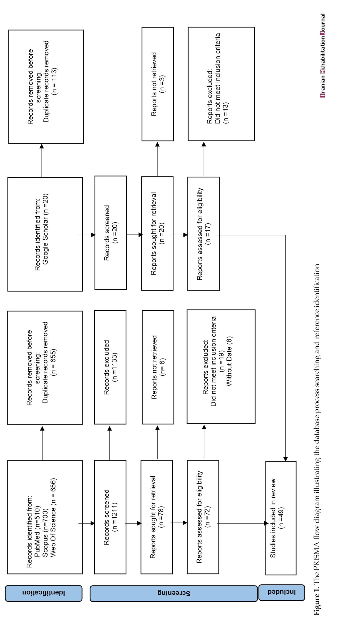
A total of 1866 articles were identified in the selected databases (Figure 1).
It is widely acknowledged that physical exercise is essential for maintaining overall health and wellbeing [1]. However, engaging in physical exercise can bring about injuries and may have some detrimental effects on overall performance of people [2]. To illustrate, injuries to the ankle and knee are frequent, particularly in activities that involve cutting and jumping movements [3]. During the landing phases of jumping exercises, substantial loads are typically placed on the leg extensor muscles, which work eccentrically to slow down the body’s downward motion and dissipate the kinetic energy generated upon landing [4]. As a consequence, studies have investigated the biomechanics and pathomechanics of landing to improve comprehension of injury mechanisms, aiming to develop strategies for prevention and treatments [5-7].
Landing is well recognized as a key movement that reflects the neuromuscular system’s capacity to control motion, especially in running and jumping activities [1, 8]. During landing, the knee plays a crucial role in absorbing impact forces by modulating muscle activity to control downward movement, while ankle dorsiflexion and hip flexion also significantly contribute to energy absorption [9]. When the lower extremity joints effectively manage descent through sagittal plane motion [10], landings tend to be safer because the ligaments responsible for lateral joint stability experience reduced loading [11]. Nevertheless, during prolonged activity, the body experiences a temporary decline in performance capacity, known as physical fatigue, which is an external factor impacting the neuromusculoskeletal system [12].
Neuromuscular control considerably contributes to maintaining dynamic joint stability and protecting the human body against various injuries [13, 14]; neuromuscular fatigue can impair this control and stability [15]. Prior studies demonstrate that neuromuscular fatigue can trigger various biomechanical alterations in the body, potentially increasing the risk of anterior cruciate ligament (ACL) injuries during landing [16, 17]. This injury may imply several impairments, including abnormal postural control, altered landing patterns, and neuromuscular deficits that can exacerbate the negative consequences of fatigue [18-20]. Yet, previous findings on this issue have some inconsistencies, and the impact of fatigue on lower limb injuries remains uncertain. For instance, in single-limb landings, researchers have observed that fatigue can lead to numerous biomechanical changes in the lower limbs, including decreased knee flexion and adduction moments [21]. Moreover, some studies have found that fatigue induces biomechanical changes that can be observed in both the lower extremity of the ACL reconstruction and the uninvolved one [18, 22].
Previous researchers have identified sagittal plane variables as factors that may contribute to the mechanism of lower extremity injuries, such as ACL [23, 24]. Research has shown that limited movement in the sagittal plane increases knee valgus angles and reduces energy absorption in the hip and knee joints among female soccer players [25]. These findings suggest that landing with limited motion in this plane could raise the risk of ACL injury. Additionally, other studies have demonstrated that reduced hip flexion and increased external knee flexion moments are associated with a higher likelihood of ACL injury [26]. It is shown that localized muscle fatigue in the lower extremity limbs has a significant association with greater alterations in postural stability in either the frontal or sagittal plane [27]. Another study suggests that the fatigue of the ankle dorsiflexors and plantar-flexors may have detrimental effects on postural stability in the sagittal plane only [28]. Moreover, lower extremity injuries can lead to alterations in angles and velocities, particularly during movements involving changes in direction, such as sidestepping, or during the landing phase of a jump [29]. Additionally, it has been observed that females have greater variation in knee joint angular velocity during the landing phase than males [30].
Despite extensive research on fatigue-induced changes in lower extremity kinematics during landing, inconsistencies persist due to methodological variations in fatigue protocols, kinematic analysis, and participant characteristics. While a recent article [31] has offered valuable insights into sex-specific responses to fatigue during landing tasks, the focus remains narrowly centered on gender-based comparisons and ACL-related outcomes. In contrast, this review expands the scope to provide a comprehensive synthesis of fatigue-induced changes in lower extremity landing kinematics across diverse fatigue protocols and study designs, regardless of sex or injury status. Our analysis emphasizes kinematic alterations as a function of fatigue, encompassing a broader spectrum of biomechanical variables. By aggregating and quantifying the impact of fatigue on lower extremity joint angles, our work provides a more comprehensive and versatile framework that can inform injury prevention, athletic training, and rehabilitation across various sports and populations. Therefore, this systematic review aimed to synthesize and gather available data on the effect of fatigue on the biomechanics of lower extremity limbs (including the hip, knee, and ankle) during landing in physically active individuals.
Materials and Methods
This review and meta-analysis study was registered prospectively in PROSPERO under the number CRD 42024502034.
Search strategy and keywords
This study used the preferred reporting items for systematic reviews and meta-analyses (PRISMA) guidelines [32]. All relevant articles were extracted using the search approach. Using a combination of phrases related to “lower extremity,” “kinematics,” “biomechanics,” “fatigue,” and “landing,” we conducted a systematic search across Scopus, Web of Science, and PubMed from the databases’ inception until June 2025 to identify relevant papers. Google Scholar was also searched. The references of pertinent papers were also carefully screened by three independent reviewers (Mohammad Salsali, Parisa Sayyadi, and Ebrahim Ebrahimi to see possibly more relevant literature and consulted with an expert (Rahman Sheikhhoseini) in the research area.
The following keyword combinations were used with the help of AND and OR operators as follows: ([Thigh OR Shank OR hip OR knee OR ankle OR foot OR feet OR “lower extremity” OR “lower limb” OR “lower-limb” OR “lower-extremity”] AND [fatigue* OR lassitude OR tiredness OR exhaustion) AND (Kinematics OR Biomechanics OR “human movement analysis” OR “motion analysis” OR “lower limb motion”] AND [jump* OR “touchdown” OR land* OR “take-off” OR Task*]).
Inclusion and exclusion criteria
A study was included in this review if it met the following criteria: 1) Examined the effect of fatigue on lower limb kinematics among physically active individuals, 2) was written in English, and 3) was published in a peer-reviewed journal. The following exclusion criteria were implemented: 1) studies were performed on individuals with neurological problems, ligament laxity, and lower extremity injury; 2) studies did not provide sufficient information for meta-analyses; and 3) studies were published as conferences, papers, abstracts, and unpublished dissertations.
Study selection
In this study, 3 authors (Mohammad Salsali, Parisa Sayyadi, and Elnaz Rajabi) independently examined and selected the articles’ titles and abstracts according to the inclusion criteria and PRISMA standard methodology, utilizing a standardized Excel data extraction sheet [33]. All human studies and trials published until the end of the search period (June 2025) were included. The supervising author (Rahman Sheikhhoseini) addressed and assessed discrepancies between the researchers. Their searched records were imported into EndNote software, version 20. This software was also used to remove duplicate articles.
Data extraction and quality assessment
Two researchers (Mohammad Salsali and Ebrahim Ebrahimi ) employed the Joanna Briggs institute (JBI) critical appraisal tools [34] to evaluate the potential for bias, selecting the specific tool according to the research design included in each study and the case-control analysis. Using a standard Excel data extraction sheet, the researchers independently collected data and subsequently compared their findings to evaluate coherence. Furthermore, the supervising author addressed and assessed any discrepancies between the researchers (Rahman Sheikhhoseini). The subsequent statistics were extracted from the included research based on the first author, year of publication, type of study, quality, sample size, subjects (including age, gender, and index data), the most important methods and tools used for data collection, and the most significant results obtained.
Data analysis
In this study, we used the comprehensive meta-analysis CMA software, version 4.0 (Biostat Inc., Englewood, New Jersey). The required data from eligible articles include the standard deviation, mean of pre- and post-tests, P values of the sample size, and mean difference. The effect size was calculated using Hedges’ g to account for differences in measurement scales. The I2 was employed to evaluate the data’s heterogeneity and estimate the heterogeneity percentage. The Funnel Plot methods were used to assess the articles’ publication bias risk. In the event of potential bias observed using this method and to investigate the extent to which the articles used in this field might affect the final results of this meta-analysis, we employed the trim-and-fill method.
Results
A total of 1866 articles were identified in the selected databases (Figure 1).
Once the data were entered into EndNote software and duplicate records were removed, 1211 articles remained. After reviewing the abstracts and titles, 72 articles were selected for further analysis. Following this, the complete text of the 53 chosen articles was carefully analysed; ultimately, 45 papers were deemed suitable for the study. Additionally, 4 studies from the Google Scholar database were included, resulting in a total of 49 studies.
Study characteristics
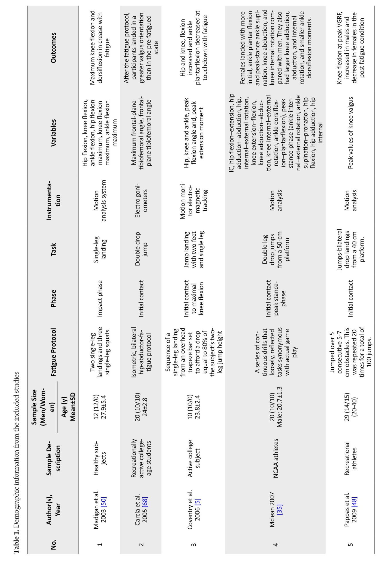
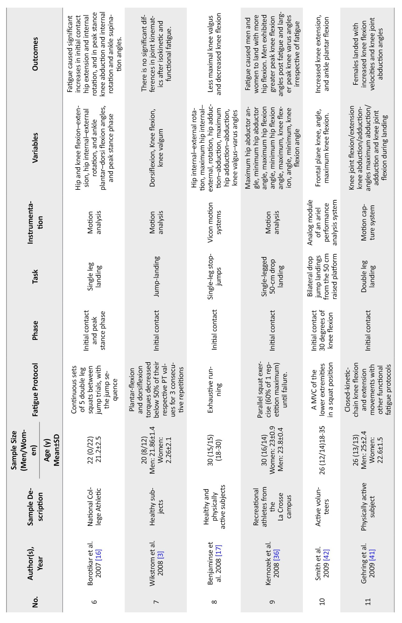
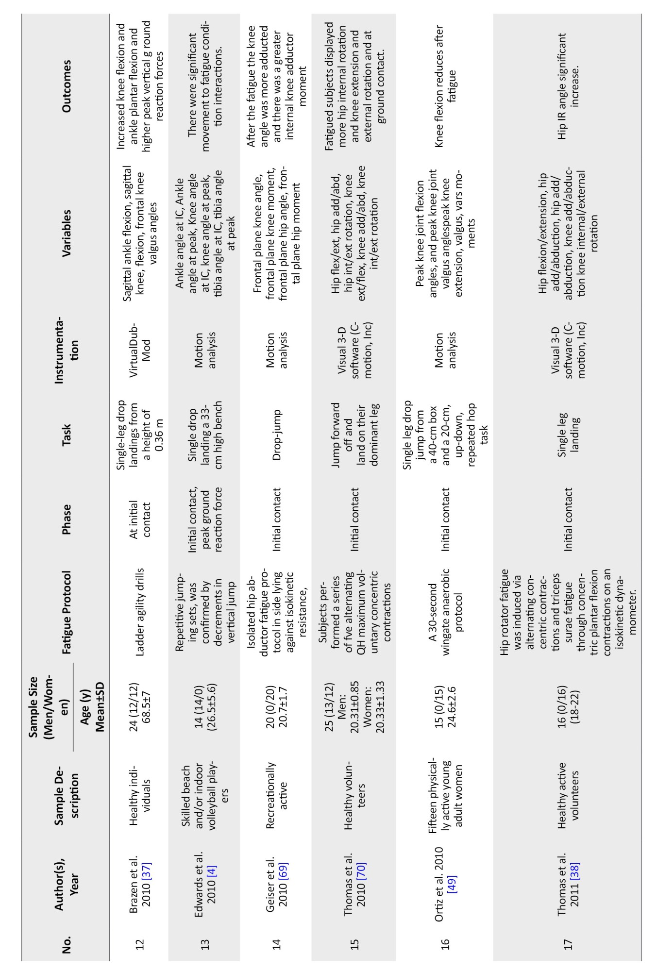
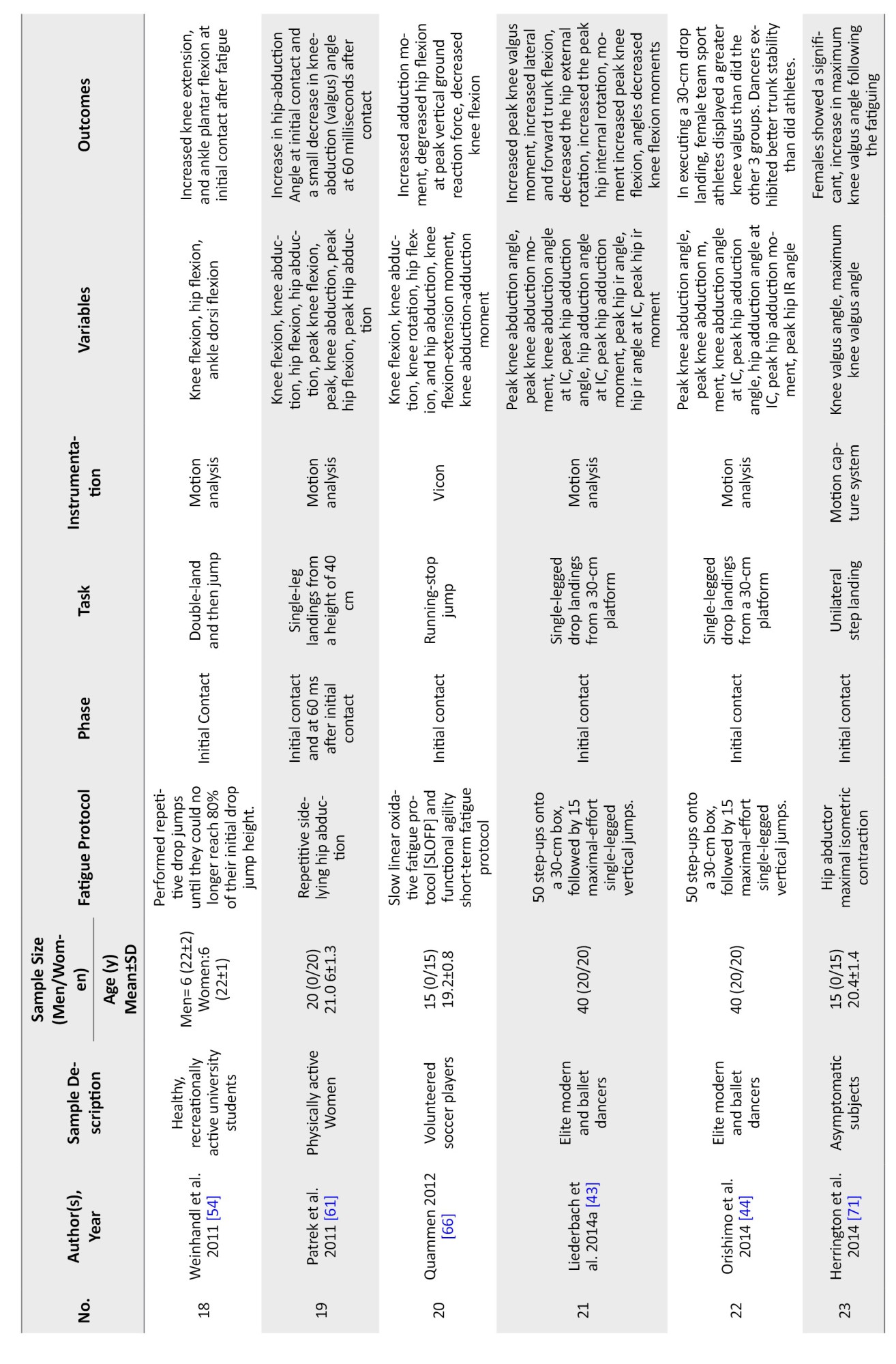
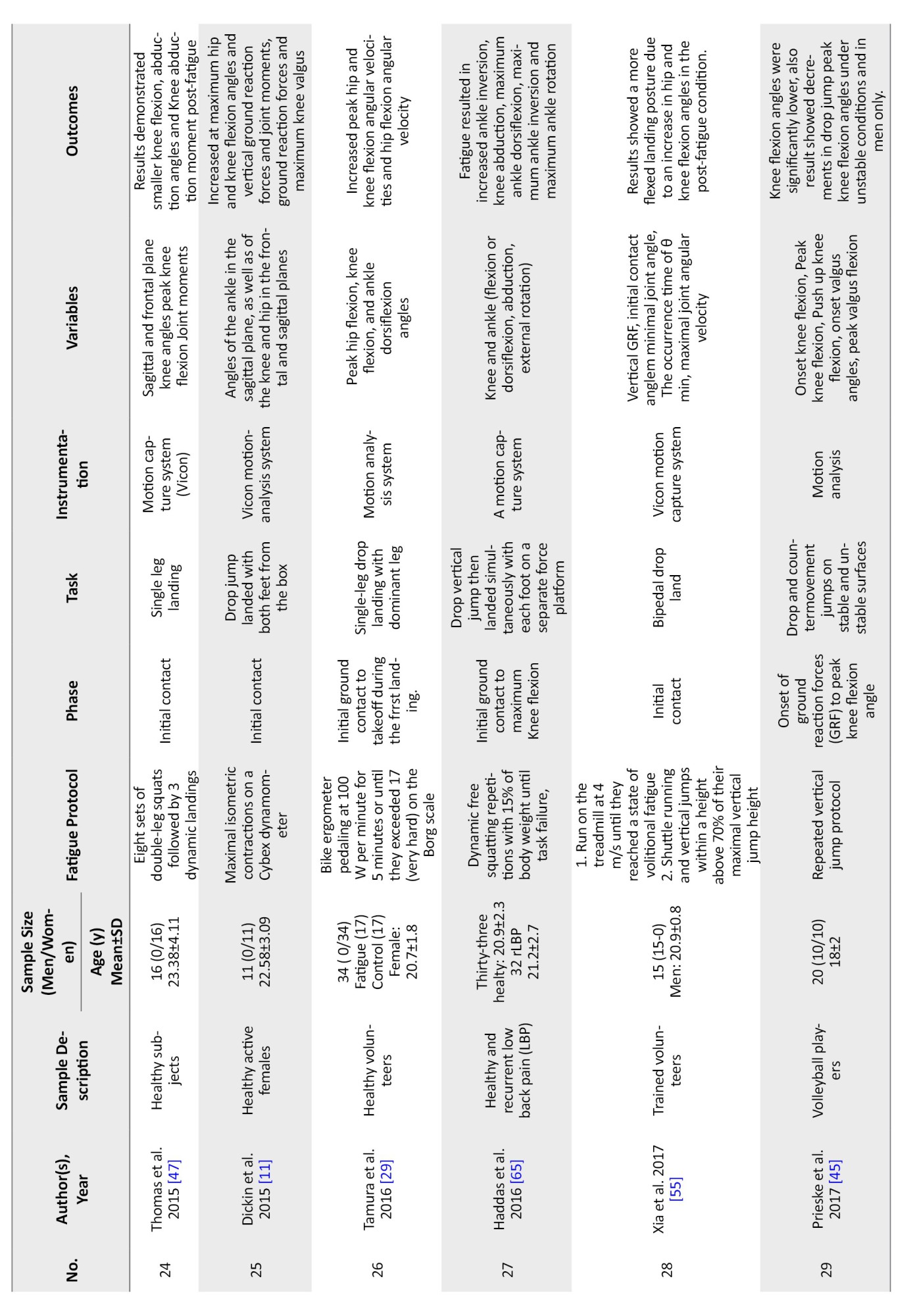
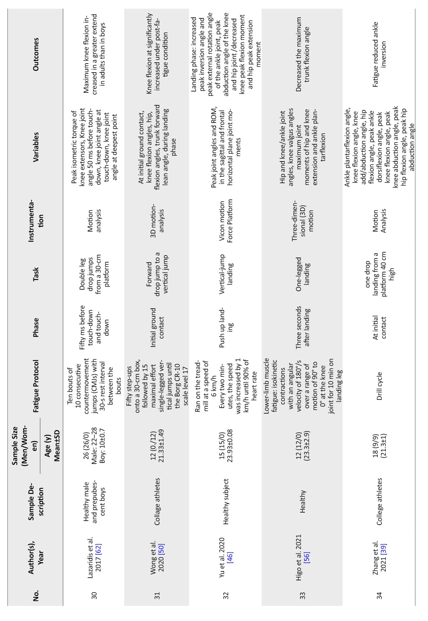
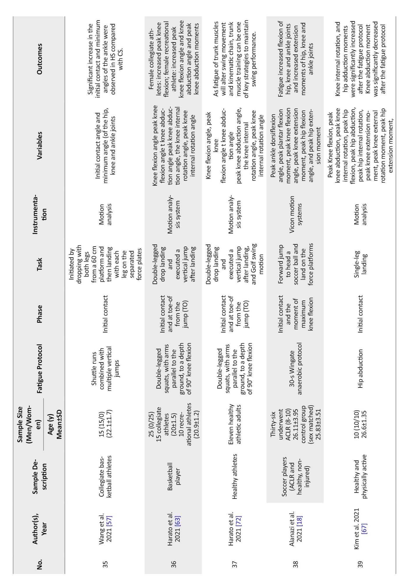
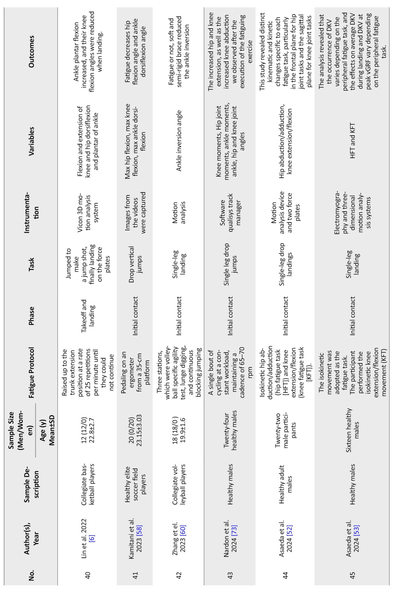
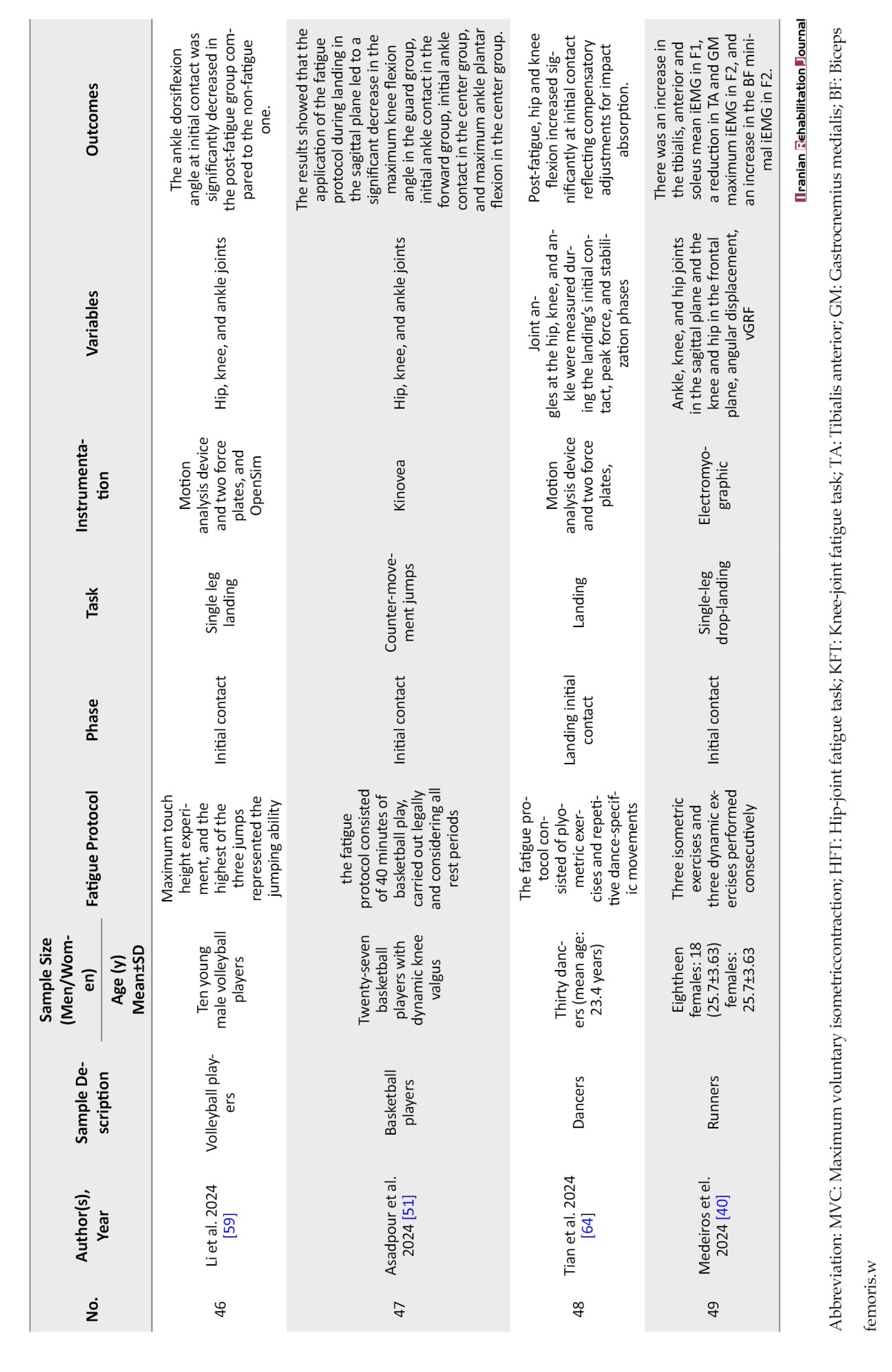
Publication dates ranged from 2003 to 2024. Of the 49 studies and 3678 participants included in this systematic review, 20 studies included only females [7, 16, 17, 29, 35-49], 21 studies included only males [4-6, 11, 18, 38, 46, 50-63], and 8 studies included both male and female [3, 48, 55, 64-67] participants. Regarding the phase of landing, 1 study used impact phase [50], 1 study used the onset of ground reaction force (GRF) to peak knee flexion angle [45], 1 study used 50 ms before touchdown and touchdown [62], 1 study used 3 s after landing [56], 1 study used take-off and landing [6], and the other studies utilized initial contact phase. Also, the instrumentation of 1 study was electro goniometers [68], an analogue module of an ariel performance analysis system [42], VirtualDubMod [37], QuickTime Player [59], Electromyography [53], Kinovea [51], and the other studies used Vicon motion capture system and force plate. Online supplemental tables contain detailed information about each included study (Table 1).
Risk of bias
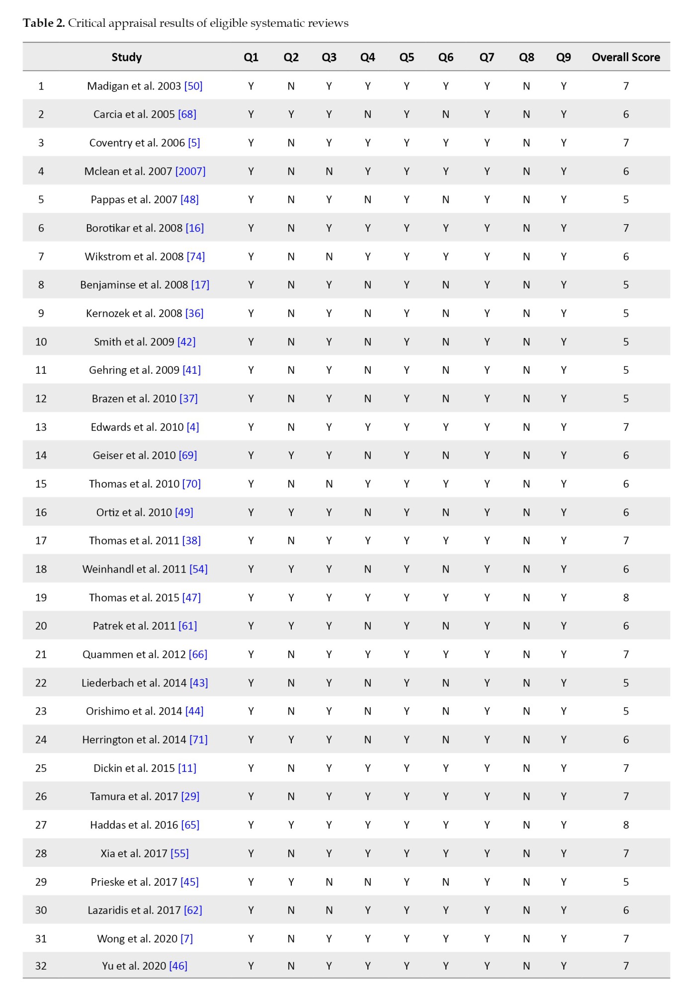
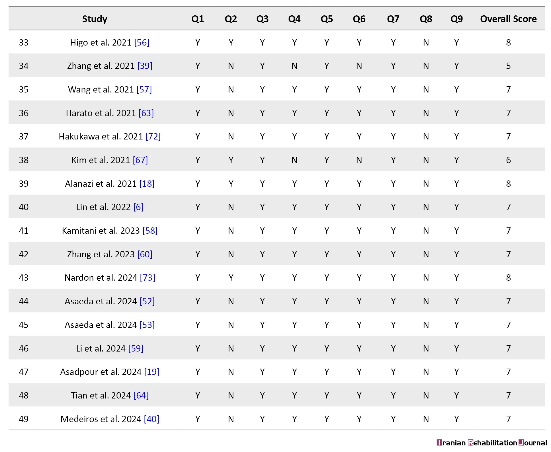
A methodological quality assessment was conducted across all included studies using a standardized checklist (JBI) comprising 9 criteria. Overall, the studies demonstrated moderate methodological quality, with quality scores ranging from 5 to 8 out of a possible 9. The most consistently met criteria were related to clarity in the study objective, the use of valid outcome measures, and the use of appropriate data collection methods. However, a notable and consistent source of bias was observed in criterion Q8 (Were outcomes measured in a reliable way?), which was not satisfied by any of the included studies, indicating a systematic gap in reporting or execution, likely associated with issues such as blinding or follow-up completeness. Additionally, variability was observed in criteria related to statistical analysis and participant selection, suggesting a potential for selection and performance bias. Despite these limitations, a substantial proportion of the studies (over 75%) met at least 6 quality criteria, supporting the overall reliability of the findings. Nonetheless, the presence of a moderate risk of bias across studies necessitates a cautious interpretation of the pooled results (Table 2).
Fatigue effects on kinematics of ankle dorsiflexion
Twenty-seven studies [3-6, 11, 16, 18, 29, 35-40, 46, 50, 51, 54-59, 63, 65, 76, 74] were included in the meta-analysis to investigate the effect of fatigue on the kinematics of ankle dorsiflexion, a key kinematic parameter of the lower extremities during landing. A total of 498 participants were involved in these studies. Forest plot analysis of the data revealed a non-significant change in ankle dorsiflexion due to fatigue effects (P=0.372; 95% CI, -0.116%, 0.309%). Assessment of heterogeneity revealed significant variability among the studies (P=0.000; I²=77.979), suggesting substantial differences in study populations, fatigue protocols, or measurement methods. To explore potential moderators, we performed meta-regression analyses for sex, but the results showed that it did not significantly impact the observed effect sizes (P>0.05). Subgroup analyses by sex (male, female, and both) also indicated no significant differences in the effect of fatigue on ankle dorsiflexion across these groups. The funnel plot showed a symmetrical distribution of studies around the pooled effect size, and Egger’s test yielded a non-significant intercept (P=0.59776).
Fatigue effects on kinematics of ankle inversion
Five eligible studies [35, 38, 46, 60, 65] have investigated the impact of fatigue on the kinematics of ankle inversion. A total of 102 participants took part in these studies. The forest plot analysis revealed that the studies showed varied but generally minor changes in ankle inversion, with a statistically significant effect across the pooled data (95% CI, 0.114%, 0.537%; P=0.003). The heterogeneity analysis indicated no significant heterogeneity among the studies (P=0.516, I²=0.000). To evaluate publication bias, we conducted Egger’s regression test, which yielded a non-significant intercept (P=0.65654).
Fatigue effects on kinematics of ankle supination
Four studies [16, 35, 46, 65] involving a total of 93 participants were analyzed to examine the effects of fatigue on the kinematics of ankle supination. The combined effect size indicated a modest and statistically non-significant shift in ankle supination/pronation following fatigue (P=0.326; 95% CI, -0.23%, 0.692%). Heterogeneity analysis confirmed considerable variation among the studies (P=0.002, I²=76.011), likely attributable to differences in study methodologies, fatigue interventions, or participant demographics. We performed Egger’s regression test to assess publication bias, which showed a non-significant intercept (P=0.84139).
Fatigue effects on kinematics of knee flexion
Forty-five studies [3-6, 11, 12, 16-18, 29, 35-39, 41-51, 53-59, 61-65, 68-70, 72, 73], including a total of 906 participants, were considered in the meta-analysis to examine how fatigue affects the kinematics of knee flexion. The forest plot analysis demonstrated a range of outcomes across the studies, with no significant overall effect found in the combined data (P=0.885, 95% CI, -0.135%, 0.156%). Analysis of heterogeneity revealed considerable differences among the studies (P=0.000, I²=72.707). We performed subgroup analyses by sex, but the factor did not significantly influence the effect sizes. To check for publication bias, we conducted Egger’s regression test, and it indicated a non-significant intercept (P=0.38787).
Fatigue effects on kinematics of knee adduction
Twenty-nine studies [3, 16, 17, 35, 36, 38-43, 45-49, 53, 54, 62, 64, 66, 68-75], with a total of 672 participants, were incorporated into the meta-analysis to explore the impact of fatigue on the kinematics of knee adduction. Data analysis using CMA and forest plot analysis demonstrated a minor and statistically non-significant difference in knee adduction/abduction following fatigue (P=0.402, 95% CI, -0.060%, 0.149%). Also, heterogeneity evaluation revealed moderate inconsistency among the studies (P=0.069, I²=28.877). Subgroup analyses based on sex revealed no significant changes in the effect sizes. Moreover, Egger’s regression test yielded a non-significant intercept (P=0.60201).
Fatigue effects on kinematics of knee internal rotation
Nine studies [16, 35, 38, 46, 53, 65, 66, 67, 70] examined the possible effects of fatigue on the kinematics of knee rotation. The total number of participants in these studies was 231. The overall pooled estimate was P=0.263 with 95% CI, -0.091%, 0.334%. Also, after examining the heterogeneity, the results showed that the heterogeneity was significant (P=0.087, I²=42.023). To examine publication bias, we conducted Egger’s regression test. It showed a non-significant intercept (P=0.93192). In addition, subgroup analysis based on sex was insignificant.
Fatigue effects on kinematics of hip flexion
Twenty-six studies [5, 6, 11, 16, 18, 29, 35, 36, 38-40, 46, 7, 50, 51, 54-59, 61, 64, 66, 67, 70], involving a total of 501 participants, were included in the meta-analysis to investigate the impact of fatigue on hip flexion. The forest plot analysis showed a range of outcomes across the studies, with no significant overall effect observed in the combined data (P=0.947, 95% CI, -0.206%, 0.192%). Heterogeneity assessment indicated significant variability among the studies (P=0.000, I²=73.481). Likewise, the subgroup analysis revealed that sex has a minimal influence on the effect sizes. We evaluated a funnel plot to examine publication bias and conducted Egger’s regression test. The funnel plot displayed a symmetrical distribution of studies around the pooled effect size, and Egger’s test yielded a non-significant intercept (P=0.44254).
Fatigue effects on kinematics of hip abduction
Seventeen studies [11, 17, 35, 36, 38, 39, 43, 44, 46, 52, 53, 61, 66, 67, 69, 70], with a total of 382 participants, were included in the meta-analysis to explore the effect of fatigue on hip abduction. The overall pooled effect size indicated a non-significant change in hip adduction/abduction following fatigue (P=0.516; 95% CI, -0.099 %, 0.197%) . Heterogeneity assessment showed significant differences among the studies (P=0.034, I²=39.998). With the moderate number of studies, we performed subgroup analyses by sex, but there was no significant influence on the effect sizes. Also, Egger’s test yielded a non-significant intercept (P=0.24714).
Fatigue effects on kinematics of hip rotation
Twelve studies [16, 17, 35, 38, 43, 44, 46, 52, 53, 67, 70, 73], comprising a total of 293 participants, were included in the meta-analysis to evaluate the influence of fatigue on hip rotation. The overall pooled effect size demonstrated a non-significant alteration in hip rotation following fatigue (P=0.760; 95% CI, -0.391%, 0.286%). Heterogeneity testing revealed significant variability among the studies (P=0.000, I²=83.642). To assess publication bias, we conducted Egger’s regression test, and it demonstrated a non-significant intercept (P=0.04839).
Discussion
The findings of this study suggest that fatigue does not substantially affect knee, hip, or ankle joint angles across the sagittal, frontal, or horizontal planes. However, a notable exception was observed in ankle inversion, where a significant effect of fatigue was detected, highlighting a potential area of concern for performance changes. These results contribute to understanding the impact of fatigue on joint kinematics during landing, providing valuable insights for strength and conditioning professionals and informing injury prevention strategies. While this study focused on kinematic variables, it is vital to recognize that changes in kinetics and muscle activity (electromyography) may be more sensitive indicators of fatigue-induced alterations. The limited impact of fatigue observed in joint angles suggests that gross kinematic changes alone may not fully show the biomechanical consequences of fatigue.
Multiple factors might have obscured significant biomechanical predisposing factors for lower extremity injuries during fatigued sessions. Enhanced muscular control, particularly eccentric control of the joint [75], and compensatory neuromuscular strategies [76] are potential mechanisms that can prevent dynamic instability in the lower limbs. A review examining fatigue-induced biomechanical changes in single-limb landings showed that vertical ground reaction forces and hip and knee joint moments were reduced after fatigue [77]. Another study by Jayalath et al. demonstrated that fatigue affects ankle biomechanics by reducing dorsiflexion from initial contact to maximum knee flexion at landing [78]. Importantly, the lack of statistically significant findings should not be misinterpreted as an absence of biomechanical or clinical relevance. Instead, the substantial variability observed across studies likely reflects subject-specific adaptation strategies in response to fatigue. Fatigue does not impact all individuals uniformly; rather, it can provoke a range of neuromuscular responses depending on each individual’s conditioning level, motor control strategies, previous injury history, and even psychological factors such as risk perception or movement confidence [79]. A review by Liu et al. demonstrates a significant change in hip external rotation and knee flexion angle after fatigue [31]. Although the study by Liu reported significant changes in knee flexion and hip external rotation angles following fatigue, their conclusions were based on a relatively limited dataset comprising only 14 included studies. Specifically, their analysis of knee flexion relied on just 10 studies, while only 3 studies contributed data on hip external rotation. In contrast, our meta-analysis incorporated a substantially larger evidence base, including 49 studies in total, with 45 studies analyzing knee flexion and 12 studies examining hip external rotation. This broader inclusion not only strengthens the statistical power and generalizability of our findings but also suggests that the significance levels observed in smaller datasets may shift when analyzed with more comprehensive data.
Additionally, the human body possesses adaptive mechanisms that can act to preserve movement patterns despite the presence of fatigue. These compensatory responses can involve neuromuscular adjustments, such as modified muscle activation patterns or increased co-contraction, which help maintain joint stability and mitigate unfavorable kinematic deviations [80]. Also, the lack of consistent directional change across studies in the meta-analysis may reflect these individualized strategies for mitigating fatigue-induced impairments, rather than a true absence of effect. Moreover, in athletic contexts, this rigid control pattern may act as a short-term protective mechanism but may also predispose individuals to overuse or load-related injuries if sustained over time [81]. These stiffening responses illustrate how motor control adjustments under fatigue can mask changes in observable kinematics [82] while substantially altering the underlying kinetic profile of movement.
Additionally, central nervous system mechanisms also play a role in compensating for fatigue. The brain may adapt motor commands to preserve task performance by reorganizing motor unit recruitment, altering movement strategies, or prioritizing stability over efficiency [83]. These central adaptations may limit the extent of observable kinematic changes, effectively masking fatigue-induced perturbations at the joint level. Thus, the absence of significant group-level kinematic alterations should not be interpreted as evidence of no fatigue effect. Instead, it may reflect a complex interplay of individualized neuromechanical strategies and central compensatory processes that act to maintain motor performance under fatigue.
A notable alteration was detected in the ankle inversion. This outcome aligns with earlier studies that associate fatigue with disruptions in neuromuscular coordination and diminished joint stability, especially in the ankle region. In addition, increased ankle inversion following fatigue is clinically significant, as it directly elevates the risk of lateral ankle sprains [84]. Also, the peroneus longus plays a vital role in resisting excessive inversion during landing; when fatigued, its delayed activation and reduced force output compromise this protective mechanism. A study by Gutierrez et al. demonstrates that fatigue can decrease the median frequency and strength of the peroneus longus, a key muscle responsible for resisting ankle inversion during dynamic tasks, such as landing [85]. In a similar vein, Delahunt et al. demonstrated that fatigue impairs sensorimotor performance, resulting in delayed neuromuscular activation and a heightened tendency toward inversion deviations, thereby increasing the risk of instability and injury [86]. Moreover, fatigue-induced alterations in ankle inversion may impact knee and hip kinematics and kinetics by upstreaming the kinetic chain. Specifically, increased ankle inversion may restrict normal subtalar pronation, reducing the foot’s ability to absorb impact forces efficiently [87]. This limited motion leads to reduced tibial internal rotation during the early stance phase, which may disrupt the natural coupling between the ankle and knee joints [88]. This is because altered ankle mechanics can change joint loading patterns and force transmission, which may put more strain on proximal joints, such as the knee and hip, and increase the risk of injury [89, 90]. Furthermore, the ankle joint, especially in the frontal plane, is particularly vulnerable to instability under fatigue conditions, largely due to its relatively smaller supporting muscles and strong dependence on reflex-based stabilization. McKeon and Hertel highlighted that fatigue can impair the finely tuned neuromuscular coordination required to maintain ankle stability during dynamic tasks, thereby increasing the likelihood of excessive inversion and potential injury [91]. In contrast to ours, Liu et al. showed no significant changes across the ankle joint after fatigue [31]. The key distinction between our study and that of Liu et al. lies in the scope of ankle joint analysis. While their meta-analysis focused solely on peak ankle dorsiflexion, it did not examine other critical kinematic parameters, such as ankle inversion. Moreover, their conclusions regarding ankle biomechanics were derived from only 3 studies, significantly limiting the generalizability of their findings. In contrast, our review incorporates 36 studies that collectively provide a far more comprehensive assessment of ankle kinematics, including inversion and other angular displacements across multiple planes of motion.
One possible explanation for the absence of significant effects on knee internal rotation and adduction is the measurement challenges. The limitations of existing motion capture methods make it particularly challenging to accurately measure knee internal rotation. Internal rotation is typically difficult for inertial measurement unit sensors to detect, and even marker-based motion capture systems like Vicon have problems because of skin artifacts, which occur when the skin moves in relation to the underlying bone [92]. Additionally, there is a lack of standardization in measurement protocols, including variations in marker placement, skeletal models, and methods for calculating joint angles (e.g. direct versus inverse kinematics models), which further complicates comparisons across studies. The small magnitude of knee internal rotation angles, coupled with relatively large errors from skin movement, may obscure true kinematic differences caused by fatigue [93]. Similar methodological errors cause some variability in knee adduction measurements, even though they are less impacted by these problems [94]. In contrast, knee flexion angles, which involve a larger range of motion, are less sensitive to marker placement errors and provide more consistent results across different laboratories [95].
Furthermore, because knee moments are less dependent on small angular changes and instead represent the pressures acting on the joint, they may offer deeper insights into fatigue-induced changes in joint loading. However, as with kinematic measurements, the comparability of results may be limited by the lack of consistency in knee moment calculations among laboratories [96]. Also, due to differences in muscle activation patterns, arthrokinematic motions (such as anterior-posterior translation), and other biomechanical considerations, the same osteokinematic motion can produce substantially different loading conditions within the knee [97]. Future research should include kinetic and electromyographic data to better understand how fatigue affects lower extremity biomechanics, as these differences might not be adequately captured by kinematic analysis alone.
Substantial heterogeneity was evident across all variables, attributed to the vast array of fatigue protocols employed in the studies, which encompassed plyometric exercises, local muscle exercises with isokinetic dynamometers, stair climbing, jumping, and treadmill activities. The diversity in exercise types and variations in intensity and duration likely contributed to the observed heterogeneity among the studies. Moreover, individual differences in fitness level, training background, and neuromuscular control strategies may contribute to the variability in responses. This finding underscores the importance of establishing standardized protocols and providing more precise descriptions of exercise characteristics in future research to enhance comparability and facilitate a more comprehensive understanding of fatigue effects. In addition, the lack of publication bias in all variables except for ankle dorsi moment indicates the plausibility of the results. The conclusions should be interpreted as context-dependent rather than universal. However, further studies are needed on this variable to confirm these findings.
Several limitations related to this review should be noted. Firstly, the study was restricted to English-language articles, potentially overlooking valuable insights from non-English publications. Secondly, the focus was solely on studies involving healthy individuals, excluding research on injured populations. Thirdly, most of the variables demonstrated substantial heterogeneity, which may have influenced the overall findings. Methodological differences in kinematic measurements, particularly for knee internal rotation and adduction, where changes in motion capture equipment, marker positioning, and computational models can introduce significant variability, may exacerbate this heterogeneity. Besides, since identical osteokinematic motions may obscure differences in muscle activation or arthrokinematic movements that influence injury risk, relying solely on kinematic data restricts the ability to capture changes in joint loading. Lastly, the study only considered research investigating the acute effect of fatigue, neglecting potential long-term effects. It is also important to recognize that most studies included in this meta-analysis examined specific, often isolated, time points during landing tasks, typically pre- and post-fatigue conditions defined by operational thresholds. This methodological constraint limits the generalizability of findings across the full temporal profile of fatigue development. These limitations necessitate careful consideration when interpreting the results and inform future research directions.
Conclusion
Contrary to common belief, fatigue does not appear to consistently alter hip and knee landing kinematics in healthy, active individuals, though it does increase ankle inversion, potentially elevating the risk of ankle sprains. Various compensatory mechanisms are employed to mitigate the effects of fatigue across different populations. Future studies are needed to examine individual differences in the kinematics of landing following a fatigue protocol.
Ethical Considerations
Compliance with ethical guidelines
There were no ethical considerations to be considered in this research.
Funding
This research did not receive any grant from funding agencies in the public, commercial, or non-profit sectors.
Authors' contributions
Study design and data collection: Mohammad Salsali, Parisa Sayyadi, Elnaz Rajabi, Rahman Sheikhhoseini, and Ebrahim Ebrahimi; Writing: Mohammad Salsali, Parisa Sayyadi, Rahman Sheikhhoseini, Ebrahim Ebrahimi, and Trent M. Guess; Final approval: All authors.
Conflict of interest
The authors declared no conflict of interest.
Study characteristics
Publication dates ranged from 2003 to 2024. Of the 49 studies and 3678 participants included in this systematic review, 20 studies included only females [7, 16, 17, 29, 35-49], 21 studies included only males [4-6, 11, 18, 38, 46, 50-63], and 8 studies included both male and female [3, 48, 55, 64-67] participants. Regarding the phase of landing, 1 study used impact phase [50], 1 study used the onset of ground reaction force (GRF) to peak knee flexion angle [45], 1 study used 50 ms before touchdown and touchdown [62], 1 study used 3 s after landing [56], 1 study used take-off and landing [6], and the other studies utilized initial contact phase. Also, the instrumentation of 1 study was electro goniometers [68], an analogue module of an ariel performance analysis system [42], VirtualDubMod [37], QuickTime Player [59], Electromyography [53], Kinovea [51], and the other studies used Vicon motion capture system and force plate. Online supplemental tables contain detailed information about each included study (Table 1).
Risk of bias
A methodological quality assessment was conducted across all included studies using a standardized checklist (JBI) comprising 9 criteria. Overall, the studies demonstrated moderate methodological quality, with quality scores ranging from 5 to 8 out of a possible 9. The most consistently met criteria were related to clarity in the study objective, the use of valid outcome measures, and the use of appropriate data collection methods. However, a notable and consistent source of bias was observed in criterion Q8 (Were outcomes measured in a reliable way?), which was not satisfied by any of the included studies, indicating a systematic gap in reporting or execution, likely associated with issues such as blinding or follow-up completeness. Additionally, variability was observed in criteria related to statistical analysis and participant selection, suggesting a potential for selection and performance bias. Despite these limitations, a substantial proportion of the studies (over 75%) met at least 6 quality criteria, supporting the overall reliability of the findings. Nonetheless, the presence of a moderate risk of bias across studies necessitates a cautious interpretation of the pooled results (Table 2).
Fatigue effects on kinematics of ankle dorsiflexion
Twenty-seven studies [3-6, 11, 16, 18, 29, 35-40, 46, 50, 51, 54-59, 63, 65, 76, 74] were included in the meta-analysis to investigate the effect of fatigue on the kinematics of ankle dorsiflexion, a key kinematic parameter of the lower extremities during landing. A total of 498 participants were involved in these studies. Forest plot analysis of the data revealed a non-significant change in ankle dorsiflexion due to fatigue effects (P=0.372; 95% CI, -0.116%, 0.309%). Assessment of heterogeneity revealed significant variability among the studies (P=0.000; I²=77.979), suggesting substantial differences in study populations, fatigue protocols, or measurement methods. To explore potential moderators, we performed meta-regression analyses for sex, but the results showed that it did not significantly impact the observed effect sizes (P>0.05). Subgroup analyses by sex (male, female, and both) also indicated no significant differences in the effect of fatigue on ankle dorsiflexion across these groups. The funnel plot showed a symmetrical distribution of studies around the pooled effect size, and Egger’s test yielded a non-significant intercept (P=0.59776).
Fatigue effects on kinematics of ankle inversion
Five eligible studies [35, 38, 46, 60, 65] have investigated the impact of fatigue on the kinematics of ankle inversion. A total of 102 participants took part in these studies. The forest plot analysis revealed that the studies showed varied but generally minor changes in ankle inversion, with a statistically significant effect across the pooled data (95% CI, 0.114%, 0.537%; P=0.003). The heterogeneity analysis indicated no significant heterogeneity among the studies (P=0.516, I²=0.000). To evaluate publication bias, we conducted Egger’s regression test, which yielded a non-significant intercept (P=0.65654).
Fatigue effects on kinematics of ankle supination
Four studies [16, 35, 46, 65] involving a total of 93 participants were analyzed to examine the effects of fatigue on the kinematics of ankle supination. The combined effect size indicated a modest and statistically non-significant shift in ankle supination/pronation following fatigue (P=0.326; 95% CI, -0.23%, 0.692%). Heterogeneity analysis confirmed considerable variation among the studies (P=0.002, I²=76.011), likely attributable to differences in study methodologies, fatigue interventions, or participant demographics. We performed Egger’s regression test to assess publication bias, which showed a non-significant intercept (P=0.84139).
Fatigue effects on kinematics of knee flexion
Forty-five studies [3-6, 11, 12, 16-18, 29, 35-39, 41-51, 53-59, 61-65, 68-70, 72, 73], including a total of 906 participants, were considered in the meta-analysis to examine how fatigue affects the kinematics of knee flexion. The forest plot analysis demonstrated a range of outcomes across the studies, with no significant overall effect found in the combined data (P=0.885, 95% CI, -0.135%, 0.156%). Analysis of heterogeneity revealed considerable differences among the studies (P=0.000, I²=72.707). We performed subgroup analyses by sex, but the factor did not significantly influence the effect sizes. To check for publication bias, we conducted Egger’s regression test, and it indicated a non-significant intercept (P=0.38787).
Fatigue effects on kinematics of knee adduction
Twenty-nine studies [3, 16, 17, 35, 36, 38-43, 45-49, 53, 54, 62, 64, 66, 68-75], with a total of 672 participants, were incorporated into the meta-analysis to explore the impact of fatigue on the kinematics of knee adduction. Data analysis using CMA and forest plot analysis demonstrated a minor and statistically non-significant difference in knee adduction/abduction following fatigue (P=0.402, 95% CI, -0.060%, 0.149%). Also, heterogeneity evaluation revealed moderate inconsistency among the studies (P=0.069, I²=28.877). Subgroup analyses based on sex revealed no significant changes in the effect sizes. Moreover, Egger’s regression test yielded a non-significant intercept (P=0.60201).
Fatigue effects on kinematics of knee internal rotation
Nine studies [16, 35, 38, 46, 53, 65, 66, 67, 70] examined the possible effects of fatigue on the kinematics of knee rotation. The total number of participants in these studies was 231. The overall pooled estimate was P=0.263 with 95% CI, -0.091%, 0.334%. Also, after examining the heterogeneity, the results showed that the heterogeneity was significant (P=0.087, I²=42.023). To examine publication bias, we conducted Egger’s regression test. It showed a non-significant intercept (P=0.93192). In addition, subgroup analysis based on sex was insignificant.
Fatigue effects on kinematics of hip flexion
Twenty-six studies [5, 6, 11, 16, 18, 29, 35, 36, 38-40, 46, 7, 50, 51, 54-59, 61, 64, 66, 67, 70], involving a total of 501 participants, were included in the meta-analysis to investigate the impact of fatigue on hip flexion. The forest plot analysis showed a range of outcomes across the studies, with no significant overall effect observed in the combined data (P=0.947, 95% CI, -0.206%, 0.192%). Heterogeneity assessment indicated significant variability among the studies (P=0.000, I²=73.481). Likewise, the subgroup analysis revealed that sex has a minimal influence on the effect sizes. We evaluated a funnel plot to examine publication bias and conducted Egger’s regression test. The funnel plot displayed a symmetrical distribution of studies around the pooled effect size, and Egger’s test yielded a non-significant intercept (P=0.44254).
Fatigue effects on kinematics of hip abduction
Seventeen studies [11, 17, 35, 36, 38, 39, 43, 44, 46, 52, 53, 61, 66, 67, 69, 70], with a total of 382 participants, were included in the meta-analysis to explore the effect of fatigue on hip abduction. The overall pooled effect size indicated a non-significant change in hip adduction/abduction following fatigue (P=0.516; 95% CI, -0.099 %, 0.197%) . Heterogeneity assessment showed significant differences among the studies (P=0.034, I²=39.998). With the moderate number of studies, we performed subgroup analyses by sex, but there was no significant influence on the effect sizes. Also, Egger’s test yielded a non-significant intercept (P=0.24714).
Fatigue effects on kinematics of hip rotation
Twelve studies [16, 17, 35, 38, 43, 44, 46, 52, 53, 67, 70, 73], comprising a total of 293 participants, were included in the meta-analysis to evaluate the influence of fatigue on hip rotation. The overall pooled effect size demonstrated a non-significant alteration in hip rotation following fatigue (P=0.760; 95% CI, -0.391%, 0.286%). Heterogeneity testing revealed significant variability among the studies (P=0.000, I²=83.642). To assess publication bias, we conducted Egger’s regression test, and it demonstrated a non-significant intercept (P=0.04839).
Discussion
The findings of this study suggest that fatigue does not substantially affect knee, hip, or ankle joint angles across the sagittal, frontal, or horizontal planes. However, a notable exception was observed in ankle inversion, where a significant effect of fatigue was detected, highlighting a potential area of concern for performance changes. These results contribute to understanding the impact of fatigue on joint kinematics during landing, providing valuable insights for strength and conditioning professionals and informing injury prevention strategies. While this study focused on kinematic variables, it is vital to recognize that changes in kinetics and muscle activity (electromyography) may be more sensitive indicators of fatigue-induced alterations. The limited impact of fatigue observed in joint angles suggests that gross kinematic changes alone may not fully show the biomechanical consequences of fatigue.
Multiple factors might have obscured significant biomechanical predisposing factors for lower extremity injuries during fatigued sessions. Enhanced muscular control, particularly eccentric control of the joint [75], and compensatory neuromuscular strategies [76] are potential mechanisms that can prevent dynamic instability in the lower limbs. A review examining fatigue-induced biomechanical changes in single-limb landings showed that vertical ground reaction forces and hip and knee joint moments were reduced after fatigue [77]. Another study by Jayalath et al. demonstrated that fatigue affects ankle biomechanics by reducing dorsiflexion from initial contact to maximum knee flexion at landing [78]. Importantly, the lack of statistically significant findings should not be misinterpreted as an absence of biomechanical or clinical relevance. Instead, the substantial variability observed across studies likely reflects subject-specific adaptation strategies in response to fatigue. Fatigue does not impact all individuals uniformly; rather, it can provoke a range of neuromuscular responses depending on each individual’s conditioning level, motor control strategies, previous injury history, and even psychological factors such as risk perception or movement confidence [79]. A review by Liu et al. demonstrates a significant change in hip external rotation and knee flexion angle after fatigue [31]. Although the study by Liu reported significant changes in knee flexion and hip external rotation angles following fatigue, their conclusions were based on a relatively limited dataset comprising only 14 included studies. Specifically, their analysis of knee flexion relied on just 10 studies, while only 3 studies contributed data on hip external rotation. In contrast, our meta-analysis incorporated a substantially larger evidence base, including 49 studies in total, with 45 studies analyzing knee flexion and 12 studies examining hip external rotation. This broader inclusion not only strengthens the statistical power and generalizability of our findings but also suggests that the significance levels observed in smaller datasets may shift when analyzed with more comprehensive data.
Additionally, the human body possesses adaptive mechanisms that can act to preserve movement patterns despite the presence of fatigue. These compensatory responses can involve neuromuscular adjustments, such as modified muscle activation patterns or increased co-contraction, which help maintain joint stability and mitigate unfavorable kinematic deviations [80]. Also, the lack of consistent directional change across studies in the meta-analysis may reflect these individualized strategies for mitigating fatigue-induced impairments, rather than a true absence of effect. Moreover, in athletic contexts, this rigid control pattern may act as a short-term protective mechanism but may also predispose individuals to overuse or load-related injuries if sustained over time [81]. These stiffening responses illustrate how motor control adjustments under fatigue can mask changes in observable kinematics [82] while substantially altering the underlying kinetic profile of movement.
Additionally, central nervous system mechanisms also play a role in compensating for fatigue. The brain may adapt motor commands to preserve task performance by reorganizing motor unit recruitment, altering movement strategies, or prioritizing stability over efficiency [83]. These central adaptations may limit the extent of observable kinematic changes, effectively masking fatigue-induced perturbations at the joint level. Thus, the absence of significant group-level kinematic alterations should not be interpreted as evidence of no fatigue effect. Instead, it may reflect a complex interplay of individualized neuromechanical strategies and central compensatory processes that act to maintain motor performance under fatigue.
A notable alteration was detected in the ankle inversion. This outcome aligns with earlier studies that associate fatigue with disruptions in neuromuscular coordination and diminished joint stability, especially in the ankle region. In addition, increased ankle inversion following fatigue is clinically significant, as it directly elevates the risk of lateral ankle sprains [84]. Also, the peroneus longus plays a vital role in resisting excessive inversion during landing; when fatigued, its delayed activation and reduced force output compromise this protective mechanism. A study by Gutierrez et al. demonstrates that fatigue can decrease the median frequency and strength of the peroneus longus, a key muscle responsible for resisting ankle inversion during dynamic tasks, such as landing [85]. In a similar vein, Delahunt et al. demonstrated that fatigue impairs sensorimotor performance, resulting in delayed neuromuscular activation and a heightened tendency toward inversion deviations, thereby increasing the risk of instability and injury [86]. Moreover, fatigue-induced alterations in ankle inversion may impact knee and hip kinematics and kinetics by upstreaming the kinetic chain. Specifically, increased ankle inversion may restrict normal subtalar pronation, reducing the foot’s ability to absorb impact forces efficiently [87]. This limited motion leads to reduced tibial internal rotation during the early stance phase, which may disrupt the natural coupling between the ankle and knee joints [88]. This is because altered ankle mechanics can change joint loading patterns and force transmission, which may put more strain on proximal joints, such as the knee and hip, and increase the risk of injury [89, 90]. Furthermore, the ankle joint, especially in the frontal plane, is particularly vulnerable to instability under fatigue conditions, largely due to its relatively smaller supporting muscles and strong dependence on reflex-based stabilization. McKeon and Hertel highlighted that fatigue can impair the finely tuned neuromuscular coordination required to maintain ankle stability during dynamic tasks, thereby increasing the likelihood of excessive inversion and potential injury [91]. In contrast to ours, Liu et al. showed no significant changes across the ankle joint after fatigue [31]. The key distinction between our study and that of Liu et al. lies in the scope of ankle joint analysis. While their meta-analysis focused solely on peak ankle dorsiflexion, it did not examine other critical kinematic parameters, such as ankle inversion. Moreover, their conclusions regarding ankle biomechanics were derived from only 3 studies, significantly limiting the generalizability of their findings. In contrast, our review incorporates 36 studies that collectively provide a far more comprehensive assessment of ankle kinematics, including inversion and other angular displacements across multiple planes of motion.
One possible explanation for the absence of significant effects on knee internal rotation and adduction is the measurement challenges. The limitations of existing motion capture methods make it particularly challenging to accurately measure knee internal rotation. Internal rotation is typically difficult for inertial measurement unit sensors to detect, and even marker-based motion capture systems like Vicon have problems because of skin artifacts, which occur when the skin moves in relation to the underlying bone [92]. Additionally, there is a lack of standardization in measurement protocols, including variations in marker placement, skeletal models, and methods for calculating joint angles (e.g. direct versus inverse kinematics models), which further complicates comparisons across studies. The small magnitude of knee internal rotation angles, coupled with relatively large errors from skin movement, may obscure true kinematic differences caused by fatigue [93]. Similar methodological errors cause some variability in knee adduction measurements, even though they are less impacted by these problems [94]. In contrast, knee flexion angles, which involve a larger range of motion, are less sensitive to marker placement errors and provide more consistent results across different laboratories [95].
Furthermore, because knee moments are less dependent on small angular changes and instead represent the pressures acting on the joint, they may offer deeper insights into fatigue-induced changes in joint loading. However, as with kinematic measurements, the comparability of results may be limited by the lack of consistency in knee moment calculations among laboratories [96]. Also, due to differences in muscle activation patterns, arthrokinematic motions (such as anterior-posterior translation), and other biomechanical considerations, the same osteokinematic motion can produce substantially different loading conditions within the knee [97]. Future research should include kinetic and electromyographic data to better understand how fatigue affects lower extremity biomechanics, as these differences might not be adequately captured by kinematic analysis alone.
Substantial heterogeneity was evident across all variables, attributed to the vast array of fatigue protocols employed in the studies, which encompassed plyometric exercises, local muscle exercises with isokinetic dynamometers, stair climbing, jumping, and treadmill activities. The diversity in exercise types and variations in intensity and duration likely contributed to the observed heterogeneity among the studies. Moreover, individual differences in fitness level, training background, and neuromuscular control strategies may contribute to the variability in responses. This finding underscores the importance of establishing standardized protocols and providing more precise descriptions of exercise characteristics in future research to enhance comparability and facilitate a more comprehensive understanding of fatigue effects. In addition, the lack of publication bias in all variables except for ankle dorsi moment indicates the plausibility of the results. The conclusions should be interpreted as context-dependent rather than universal. However, further studies are needed on this variable to confirm these findings.
Several limitations related to this review should be noted. Firstly, the study was restricted to English-language articles, potentially overlooking valuable insights from non-English publications. Secondly, the focus was solely on studies involving healthy individuals, excluding research on injured populations. Thirdly, most of the variables demonstrated substantial heterogeneity, which may have influenced the overall findings. Methodological differences in kinematic measurements, particularly for knee internal rotation and adduction, where changes in motion capture equipment, marker positioning, and computational models can introduce significant variability, may exacerbate this heterogeneity. Besides, since identical osteokinematic motions may obscure differences in muscle activation or arthrokinematic movements that influence injury risk, relying solely on kinematic data restricts the ability to capture changes in joint loading. Lastly, the study only considered research investigating the acute effect of fatigue, neglecting potential long-term effects. It is also important to recognize that most studies included in this meta-analysis examined specific, often isolated, time points during landing tasks, typically pre- and post-fatigue conditions defined by operational thresholds. This methodological constraint limits the generalizability of findings across the full temporal profile of fatigue development. These limitations necessitate careful consideration when interpreting the results and inform future research directions.
Conclusion
Contrary to common belief, fatigue does not appear to consistently alter hip and knee landing kinematics in healthy, active individuals, though it does increase ankle inversion, potentially elevating the risk of ankle sprains. Various compensatory mechanisms are employed to mitigate the effects of fatigue across different populations. Future studies are needed to examine individual differences in the kinematics of landing following a fatigue protocol.
Ethical Considerations
Compliance with ethical guidelines
There were no ethical considerations to be considered in this research.
Funding
This research did not receive any grant from funding agencies in the public, commercial, or non-profit sectors.
Authors' contributions
Study design and data collection: Mohammad Salsali, Parisa Sayyadi, Elnaz Rajabi, Rahman Sheikhhoseini, and Ebrahim Ebrahimi; Writing: Mohammad Salsali, Parisa Sayyadi, Rahman Sheikhhoseini, Ebrahim Ebrahimi, and Trent M. Guess; Final approval: All authors.
Conflict of interest
The authors declared no conflict of interest.
References
- Xia R, Zhang XN, FU WJ. Effects of two fatigue protocols on lower extremity kinematics and time/frequency-domain characteristics of impact forces during drop landing. Journal of Medical Biomechanics. 2017; 427-35. [DOI:10.1155/2017/5690519] [PMID]
- Chéron C, Le Scanff C, Leboeuf-Yde C. Association between sports type and overuse injuries of extremities in adults: A systematic review. Chiropractic & Manual Therapies. 2017; 25:1-10. [DOI:10.1186/s12998-017-0135-1] [PMID]
- Wikstrom EA, Powers ME, Tillman MD. Dynamic stabilization time after isokinetic and functional fatigue. Journal of Athletic Training. 2004; 39(3):247. [PMID]
- Edwards S, Steele JR, McGhee D. Does a drop landing represent a whole skill landing and is this moderated by fatigue? Scandinavian Journal of Medicine & Science in Sports. 2010; 20(3):516-23. [DOI:10.1111/j.1600-0838.2009.00964.x] [PMID]
- Coventry E, O’Connor KM, Hart BA, Earl JE, Ebersole KT. The effect of lower extremity fatigue on shock attenuation during single-leg landing. Clinical Biomechanics. 2006; 21(10):1090-7. [DOI:10.1016/j.clinbiomech.2006.07.004] [PMID]
- Lin HT, Kuo WC, Chen Y, Lo TY, Li YI, Chang JH. Effects of fatigue in lower back muscles on basketball jump shots and landings. Physical Activity and Health. 2022; 6(1). [DOI:10.5334/paah.199]
- Wong TL, Huang CF, Chen PC. Effects of lower extremity muscle fatigue on knee loading during a forward drop jump to a vertical jump in female athletes. Journal of Human Kinetics. 2020; 72:5-13. [DOI:10.2478/hukin-2019-0122] [PMID]
- Fu W, Liu Y, Zhang S. Effects of footwear on impact forces and soft tissue vibrations during drop jumps and unanticipated drop landings. International Journal of Sports Medicine. 2013; 34(06):477-83. [DOI:10.1055/s-0032-1327696] [PMID]
- Laible C, Sherman OH. Risk factors and prevention strategies of non-contact anterior cruciate ligament injuries. Bulletin of the Hospital for Joint Diseases. 2015; 73(1):70-5. [Link]
- Sedaghati P, Alghosi M, Hosseini F, Ghafouri M, Fallahi Farrash F, Parvaneh Sarand A. Correlation between sagittal plane curvature of the upright human spine and postural stability in adults. Physical Treatments-Specific Physical Therapy. 2024; 14(4):303-10. [DOI:10.32598/ptj.14.4.584.2]
- Dickin DC, Johann E, Wang H, Popp JK. Combined effects of drop height and fatigue on landing mechanics in active females. Journal of Applied Biomechanics. 2015; 31(4):237-43. [DOI:10.1123/jab.2014-0190] [PMID]
- Chappell JD, Herman DC, Knight BS, Kirkendall DT, Garrett WE, Yu B. Effect of fatigue on knee kinetics and kinematics in stop-jump tasks. The American Journal of Sports Medicine. 2005; 33(7):1022-9. [DOI:10.1177/0363546504273047] [PMID]
- Lephart SM, Henry TJ. Functional rehabilitation for the upper and lower extremity. Orthopedic Clinics of North America. 1995; 26(3):579-92. [DOI:10.1016/S0030-5898(20)32017-4]
- Parsa R, Jafarnezhadgero AA, Moradzadeh N, Piri E. Effect of double-density insoles on muscle activity frequency during running and side cutting in adolescent volleyball players. Physical Treatments-Specific Physical Therapy. 2025; 15(4):265-74. [DOI:10.32598/ptj.15.4.348.16]
- Carpenter JE, Blasier RB, Pellizzon GG. The effects of muscle fatigue on shoulder joint position sense. The American Journal of Sports Medicine. 1998; 26(2):262-5. [DOI:10.1177/03635465980260021701] [PMID]
- Borotikar BS, Newcomer R, Koppes R, McLean SG. Combined effects of fatigue and decision making on female lower limb landing postures: Central and peripheral contributions to ACL injury risk. Clinical Biomechanics. 2008; 23(1):81-92. [DOI:10.1016/j.clinbiomech.2007.08.008] [PMID]
- Benjaminse A, Habu A, Sell TC, Abt JP, Fu FH, Myers JB, et al. Fatigue alters lower extremity kinematics during a single-leg stop-jump task. Knee Surgery, Sports Traumatology, Arthroscopy. 2008; 16:400-7. [DOI:10.1007/s00167-007-0432-7] [PMID]
- Alanazi AD, Mitchell K, Roddey T, Alenazi AM, Alzhrani MM, Almansour AM, et al. The effects of a high-intensity exercise bout on landing biomechanics post anterior cruciate ligament reconstruction: A quasi-experimental study. BMC Sports Science, Medicine and Rehabilitation. 2021; 13:1-8. [DOI:10.1186/s13102-021-00263-7] [PMID]
- Asadpour R, Norasteh AA. Comparing the knee valgus angle and pattern of dominant and non-dominant feet in basketball players with dynamic knee valgus after fatigue. Physical Treatments-Specific Physical Therapy. 2024; 14(4):321-30. [DOI:10.32598/ptj.14.4.604.2]
- Sheikhalizade H, Imani F, Jafarnezhadgero A, Imani Brouj S. Sand exercises altered muscular frequency content in individuals with anterior cruciate ligament reconstruction and pronated feet during walking. Physical Treatments-Specific Physical Therapy. 2024; 14(2):93-100. [DOI:10.32598/ptj.14.2.348.9]
- Webster KE, Santamaria LJ, McClelland JA, Feller JA. Effect of fatigue on landing biomechanics after anterior cruciate ligament reconstruction surgery. Medicine and Science in Sports and Exercise. 2012; 44(5):910-6. [DOI:10.1249/MSS.0b013e31823fe28d] [PMID]
- Dalton EC, Pfile KR, Weniger GR, Ingersoll CD, Herman D, Hart JM. Neuromuscular changes after aerobic exercise in people with anterior cruciate ligament–reconstructed knees. Journal of Athletic Training. 2011; 46(5):476-83. [DOI:10.4085/1062-6050-46.5.476] [PMID]
- DeMorat G, Weinhold P, Blackburn T, Chudik S, Garrett W. Aggressive quadriceps loading can induce noncontact anterior cruciate ligament injury. The American Journal of Sports Medicine. 2004; 32(2):477-83. [DOI:10.1177/0363546503258928] [PMID]
- Weinhandl JT, Earl-Boehm JE, Ebersole KT, Huddleston WE, Armstrong BS, O’Connor KM. Reduced hamstring strength increases anterior cruciate ligament loading during anticipated sidestep cutting. Clinical Biomechanics. 2014; 29(7):752-9. [DOI:10.1016/j.clinbiomech.2014.05.013] [PMID]
- Pollard CD, Sigward SM, Powers CM. Limited hip and knee flexion during landing is associated with increased frontal plane knee motion and moments. Clinical Biomechanics. 2010; 25(2):142-6. [DOI:10.1016/j.clinbiomech.2009.10.005] [PMID]
- Leppänen M, Pasanen K, Krosshaug T, Kannus P, Vasankari T, Kujala UM, et al. Sagittal plane hip, knee, and ankle biomechanics and the risk of anterior cruciate ligament injury: A prospective study. Orthopaedic Journal of Sports Medicine. 2017; 5(12):2325967117745487. [DOI:10.1177/2325967117745487] [PMID]
- Salavati M, Moghadam M, Ebrahimi I, Arab AM. Changes in postural stability with fatigue of lower extremity frontal and sagittal plane movers. Gait & Posture. 2007; 26(2):214-8. [DOI:10.1016/j.gaitpost.2006.09.001] [PMID]
- Gribble PA, Hertel J. Effect of hip and ankle muscle fatigue on unipedal postural control. Journal of Electromyography and Kinesiology. 2004; 14(6):641-6. [DOI:10.1016/j.jelekin.2004.05.001] [PMID]
- Tamura A, Akasaka K, Otsudo T, Shiozawa J, Toda Y, Yamada K. Fatigue influences lower extremity angular velocities during a single-leg drop vertical jump. Journal of Physical Therapy Science. 2017; 29(3):498-504. [DOI:10.1589/jpts.29.498] [PMID]
- Yu B, Lin CF, Garrett WE. Lower extremity biomechanics during the landing of a stop-jump task. Clinical Biomechanics. 2006; 21(3):297-305. [DOI:10.1016/j.clinbiomech.2005.11.003] [PMID]
- Liu C, Peng W, Qu W, Zhang Z, Sun J, He J, et al. Gender differences in the impact of fatigue on lower limb landing biomechanics and their association with anterior cruciate ligament (ACL) injuries: A systematic review and meta-analysis. Plos One. 2025; 20(5):e0321925. [DOI:10.1371/journal.pone.0321925] [PMID]
- Parums DV. Editorial: Review articles, systematic reviews, meta-analysis, and the updated preferred reporting items for systematic reviews and meta-analyses (PRISMA) 2020 guidelines. Medical Science Monitor. 2021; 27:e934475. [DOI:10.12659/MSM.934475]
- Page MJ, McKenzie JE, Bossuyt PM, Boutron I, Hoffmann TC, Mulrow CD, et al. The PRISMA 2020 statement: An updated guideline for reporting systematic reviews. BMJ. 2021; 372. [DOI:10.1136/bmj.n71] [PMID]
- Crommert ME, Ekblom MM, Thorstensson A. Activation of transversus abdominis varies with postural demand in standing. Gait & Posture. 2011; 33(3):473-7. [DOI:10.1016/j.gaitpost.2010.12.028] [PMID]
- Mclean SG, Fellin RE, Suedekum N, Calabrese G, Passerallo A, Joy S. Impact of fatigue on gender-based high-risk landing strategies. Medicine and Science in Sports and Exercise. 2007; 39(3):502-14. [DOI:10.1249/mss.0b013e3180d47f0] [PMID]
- Kernozek TW, Torry MR, Iwasaki M. Gender differences in lower extremity landing mechanics caused by neuromuscular fatigue. The American Journal of Sports Medicine. 2008; 36(3):554-65. [DOI:10.1177/0363546507308934] [PMID]
- Brazen DM, Todd MK, Ambegaonkar JP, Wunderlich R, Peterson C. The effect of fatigue on landing biomechanics in single-leg drop landings. Clinical Journal of Sport Medicine. 2010; 20(4):286-92. [DOI:10.1097/JSM.0b013e3181e8f7dc] [PMID]
- Thomas AC, Palmieri-Smith RM, McLean S. Isolated hip and ankle fatigue are unlikely risk factors for anterior cruciate ligament injury. Scandinavian Journal of Medicine & Science in Sports. 2011; 21(3):359-68. [DOI:10.1111/j.1600-0838.2009.01076.x] [PMID]
- Zhang Q, Ruan M, Singh NB, Huang L, Zhang X, Wu X. Progression of fatigue modifies primary contributors to ground reaction forces during drop landing. Journal of Human Kinetics. 2021; 76:161. [DOI:10.2478/hukin-2021-0052] [PMID]
- de Souza Medeiros V, da Costa FD, da Costa Curi GOB, Barbosa VD, Dionisio VC. Trunk fatigue effect on lower limb kinematic and electromyographic patterns during the single-leg drop-landing test in female novice runners. Observatório De La Economía Latinoamericana. 2024; 22(12):e8273-e. [DOI:10.55905/oelv22n12-159]
- Gehring D, Melnyk M, Gollhofer A. Gender and fatigue have influence on knee joint control strategies during landing. Clinical Biomechanics. 2009; 24(1):82-7. [DOI:10.1016/j.clinbiomech.2008.07.005] [PMID]
- Smith MP, Sizer PS, James CR. Effects of fatigue on frontal plane knee motion, muscle activity, and ground reaction forces in men and women during landing. Journal of Sports Science & Medicine. 2009; 8(3):419. [PMID]
- Liederbach M, Kremenic IJ, Orishimo KF, Pappas E, Hagins M. Comparison of landing biomechanics between male and female dancers and athletes, part 2: Influence of fatigue and implications for anterior cruciate ligament injury. The American Journal of Sports Medicine. 2014; 42(5):1089-95. [DOI:10.1177/0363546514524525] [PMID]
- Orishimo KF, Liederbach M, Kremenic IJ, Hagins M, Pappas E. Comparison of landing biomechanics between male and female dancers and athletes, part 1: Influence of sex on risk of anterior cruciate ligament injury. The American Journal of Sports Medicine. 2014; 42(5):1082-8. [DOI:10.1177/0363546514523928] [PMID]
- Prieske O, Demps M, Lesinski M, Granacher U. Combined effects of fatigue and surface instability on jump biomechanics in elite athletes. International Journal of Sports Medicine. 2017; 38(10):781-90. [DOI:10.1055/s-0043-111894] [PMID]
- Yu P, Gong Z, Meng Y, Baker JS, István B, Gu Y. The acute influence of running-induced fatigue on the performance and biomechanics of a countermovement jump. Applied Sciences. 2020; 10(12):4319. [DOI:10.3390/app10124319]
- Thomas AC, Lepley LK, Wojtys EM, McLean SG, Palmieri-Smith RM. Effects of neuromuscular fatigue on quadriceps strength and activation and knee biomechanics in individuals post-anterior cruciate ligament reconstruction and healthy adults. Journal of Orthopaedic & Sports Physical Therapy. 2015; 45(12):1042-50. [DOI:10.2519/jospt.2015.5785] [PMID]
- Pappas E, Hagins M, Sheikhzadeh A, Nordin M, Rose D. Peak biomechanical variables during bilateral drop landings: comparisons between sex (female/male) and fatigue (pre-fatigue/post-fatigue). North American Journal of Sports Physical Therapy. 2009; 4(2):83. [PMID]
- Ortiz A, Olson SL, Etnyre B, Trudelle-Jackson EE, Bartlett W, Venegas-Rios HL. Fatigue effects on knee joint stability during two jump tasks in women. Journal of Strength and Conditioning Research. 2010; 24(4):1019-27. [DOI:10.1519/JSC.0b013e3181c7c5d4] [PMID]
- Madigan ML, Pidcoe PE. Changes in landing biomechanics during a fatiguing landing activity. Journal of Electromyography and Kinesiology. 2003; 13(5):491-8. [DOI:10.1016/S1050-6411(03)00037-3] [PMID]
- Asadpour R, Norasteh AA. The effect of a fatigue program on the kinematics of lower limb joints in basketball players with dynamic knee valgus pattern in various positions. Journal of Advanced Sport Technology. 2024; 8(2):27-44. [DOI:10.32598/ptj.14.2.604.1]
- Asaeda M, Hirata K, Ohnishi T, Ito H, Miyahara S, Fukuhara K, et al. Time course of biomechanics during jump landing before and after two different fatigue tasks. Asia-Pacific Journal of Sports Medicine, Arthroscopy, Rehabilitation and Technology. 2024; 37:40-6. [DOI:10.1016/j.asmart.2024.06.002] [PMID]
- Asaeda M, Hirata K, Ohnishi T, Ito H, Miyahara S, Mikami Y. Differences in lower-limb biomechanics during single-leg landing considering two peripheral fatigue tasks. Plos One. 2024; 19(4):e0297910. [DOI:10.1371/journal.pone.0297910] [PMID]
- Weinhandl JT, Smith JD, Dugan EL. The effects of repetitive drop jumps on impact phase joint kinematics and kinetics. Journal of Applied Biomechanics. 2011; 27(2):108-15. [DOI:10.1123/jab.27.2.108] [PMID]
- Xia R, Zhang X, Wang X, Sun X, Fu W. Effects of two fatigue protocols on impact forces and lower extremity kinematics during drop landings: implications for noncontact anterior cruciate ligament injury. Journal of Healthcare Engineering. 2017; 2017(1):5690519. [DOI:10.1155/2017/5690519] [PMID]
- Higo Y, Kuruma H. Effects of lower-limb muscle fatigue, cardiopulmonary fatigue, and brain fatiguetasks on one-legged landing motion. Physical Therapy Research. 2021; 24(3):264-71. [DOI:10.1298/ptr.E10104] [PMID]
- Wang X, Deng L, Lam WK, Yang Y, Zhang X, Fu W. Wearing cushioning shoes reduce load rates more effectively in post-fatigue than in pre-fatigue during landings. Biology. 2021; 10(10):962. [DOI:10.3390/biology10100962] [PMID]
- Kamitani A, Hara K, Fujii Y, Yoshida S. Landing posture in elite female athletes during a drop vertical jump before and after a high-intensity ergometer fatigue protocol: A study of 20 Japanese women’s soccer league players. Orthopaedic Journal of Sports Medicine. 2023; 11(6):23259671231171859. [DOI:10.1177/23259671231171859] [PMID]
- Li T, Kapilevich LV, Chen J. Effects of mild fatigue on biomechanics of single leg landing in young male volleyball players. Sensors. 2024; 24(21):6811. [DOI:10.3390/s24216811] [PMID]
- Zhang Z, Zhang M. Effect of different ankle braces on lower extremity kinematics and kinetics following special-induced fatigue for volleyball players with functional ankle instability. Heliyon. 2023; 9(6):e16380. [DOI:10.1016/j.heliyon.2023.e16380] [PMID]
- Patrek MF, Kernozek TW, Willson JD, Wright GA, Doberstein ST. Hip-abductor fatigue and single-leg landing mechanics in women athletes. Journal of Athletic Training. 2011; 46(1):31-42. [DOI:10.4085/1062-6050-46.1.31] [PMID]
- Lazaridis S, Patikas DA, Bassa E, Tsatalas T, Hatzikotoulas K, Ftikas C, et al. The acute effects of an intense stretch-shortening cycle fatigue protocol on the neuromechanical parameters of lower limbs in men and prepubescent boys. Journal of Sports Sciences. 2018; 36(2):131-9. [DOI:10.1080/02640414.2017.1287932] [PMID]
- Harato K, Morishige Y, Niki Y, Kobayashi S, Nagura T. Fatigue and recovery have different effects on knee biomechanics of drop vertical jump between female collegiate and recreational athletes. Journal of Orthopaedic Surgery and Research. 2021; 16(1):739. [DOI:10.1186/s13018-021-02893-6] [PMID]
- Tian C, Wang Y, Zhang D. Comprehensive assessment of lower limb alignment and forces during dance landings under fatigue. Molecular & Cellular Biomechanics. 2024; 21(4):531. [DOI:10.62617/mcb531]
- Haddas R, Sawyer SF, Sizer PS, Brooks T, Chyu MC, James CR. Effects of volitional spine stabilization and lower-extremity fatigue on the knee and ankle during landing performance in a population with recurrent low back pain. Journal of Sport Rehabilitation. 2017; 26(5):329-38. [DOI:10.1123/jsr.2015-0171] [PMID]
- Quammen D, Cortes N, Van Lunen BL, Lucci S, Ringleb SI, Onate J. Two different fatigue protocols and lower extremity motion patterns during a stop-jump task. Journal of Athletic Training. 2012;47(1):32-41. [DOI:10.4085/1062-6050-47.1.32] [PMID]
- Kim N, Lee SY, Lee SC, Rosen AB, Grindstaff TL, Knarr BA. Effect of isolated hip abductor fatigue on single-leg landing mechanics and simulated ACL loading. Knee. 2021; 31:118-26. [DOI:10.1016/j.knee.2021.05.007] [PMID]
- Carcia C, Eggen J, Shultz S. Hip-abductor fatigue, frontal-plane landing angle, and excursion during a drop jump. Journal of Sport Rehabilitation. 2005; 14(4):321-31. [DOI:10.1123/jsr.14.4.321]
- Geiser CF, O’Connor KM, Earl JE. Effects of isolated hip abductor fatigue on frontal plane knee mechanics. Medicine & Science in Sports & Exercise. 2010; 42(3):535-45. [DOI:10.1249/MSS.0b013e3181b7b227] [PMID]
- Thomas AC, McLean SG, Palmieri-Smith RM. Quadriceps and hamstrings fatigue alters hip and knee mechanics. Journal of Applied Biomechanics. 2010; 26(2):159-70. [DOI:10.1123/jab.26.2.159] [PMID]
- Herrington L. The effect of hip abductor muscle fatigue on frontal plane knee projection angle during step landing. International Journal of Athletic Therapy and Training. 2014; 19(4):38-43. [DOI:10.1123/ijatt.2014-0003]
- Hakukawa S, Harato K, Morita E, Nishizawa K, Kobayashi S, Niki Y, et al. Biomechanical correlation between trunk and foot kinematics during golf swing movement before and after fatigue. 2021. [DOI:10.21203/rs.3.rs-651875/v1]
- Nardon M, Ferri U, Caffi G, Bartesaghi M, Perin C, Zaza A, et al. Kinematics but not kinetics alterations to single-leg drop jump movements following a subject-tailored fatiguing protocol suggest an increased risk of ACL injury. Frontiers in Sports and Active Living. 2024; 6:1418598. [DOI:10.3389/fspor.2024.1418598] [PMID]
- Wikstrom EA. Functional VS isokinetic fatigue protocol: Effects on time to stabilization, peak vertical ground reaction forces, and joint kinematics in jump landing [doctoral dissertation]. Gainesville: University of Florida; 2003. [Link]
- Fagenbaum R, Darling WG. Jump landing strategies in male and female college athletes and the implications of such strategies for anterior cruciate ligament injury. The American Journal of Sports Medicine. 2003; 31(2):233-40. [DOI:10.1177/03635465030310021301] [PMID]
- Rodacki ALF, Fowler NE, Bennett SJ. Vertical jump coordination: fatigue effects. Medicine & Science in Sports & Exercise. 2002; 34(1):105-16. [DOI:10.1097/00005768-200201000-00017] [PMID]
- Santamaria LJ, Webster KE. The effect of fatigue on lower-limb biomechanics during single-limb landings: A systematic review. Journal of Orthopaedic & Sports Physical Therapy. 2010; 40(8):464-73. [DOI:10.2519/jospt.2010.3295] [PMID]
- Jayalath JLR, de Noronha M, Weerakkody N, Bini R. Effects of fatigue on ankle biomechanics during jumps: A systematic review. Journal of Electromyography and Kinesiology. 2018; 42:81-91. [DOI:10.1016/j.jelekin.2018.06.012] [PMID]
- Granacher U, Lesinski M, Büsch D, Muehlbauer T, Prieske O, Puta C, et al. Effects of resistance training in youth athletes on muscular fitness and athletic performance: A conceptual model for long-term athlete development. Frontiers in Physiology. 2016; 7:164. [DOI:10.3389/fphys.2016.00164] [PMID]
- Enoka RM, Duchateau J. Muscle fatigue: What, why and how it influences muscle function. The Journal of Physiology. 2008; 586(1):11-23. [DOI:10.1113/jphysiol.2007.139477] [PMID]
- Swafford A, Navalta J, Silvernail JF, Lough N, Mercer J. Physiological and biomechanical responses to indoor cycling with and without the ability to sway. International Journal of Exercise Science. 2025; 18(7):531-42. [DOI:10.70252/FQRW7720] [PMID]
- Vila-Chã C, Falla D, Correia MV, Farina D. Adjustments in motor unit properties during fatiguing contractions after training. Medicine & Science in Sports & Exercise. 2012; 44(4):616-24. [DOI:10.1249/MSS.0b013e318235d81d] [PMID]
- Millet GY, Lepers R. Alterations of neuromuscular function after prolonged running, cycling and skiing exercises. Sports Medicine. 2004; 34(2):105-16. [DOI:10.2165/00007256-200434020-00004] [PMID]
- Delahunt E, Remus A. Risk factors for lateral ankle sprains and chronic ankle instability. Journal of Athletic Training. 2019; 54(6):611-6. [DOI:10.4085/1062-6050-44-18] [PMID]
- Gutierrez GM, Jackson ND, Dorr KA, Margiotta SE, Kaminski TW. Effect of fatigue on neuromuscular function at the ankle. Journal of Sport Rehabilitation. 2007; 16(4):295-306. [DOI:10.1123/jsr.16.4.295] [PMID]
- Delahunt E, Monaghan K, Caulfield B. Changes in lower limb kinematics, kinetics, and muscle activity in subjects with functional instability of the ankle joint during a single leg drop jump. Journal of Orthopaedic Research. 2006; 24(10):1991-2000. [DOI:10.1002/jor.20235] [PMID]
- Medina McKeon JM, Hoch MC. The ankle-joint complex: A kinesiologic approach to lateral ankle sprains. Journal of Athletic Training. 2019; 54(6):589-602. [DOI:10.4085/1062-6050-472-17] [PMID]
- Rodrigues P, Chang R, Tenbroek T, Emmerik R, Hamill J. Evaluating the coupling between foot pronation and tibial internal rotation continuously using vector coding. Journal of Applied Biomechanics. 2014; 31. [DOI:10.1123/JAB.2014-0067] [PMID]
- Hewett TE, Myer GD, Ford KR, Heidt Jr RS, Colosimo AJ, McLean SG, et al. Biomechanical measures of neuromuscular control and valgus loading of the knee predict anterior cruciate ligament injury risk in female athletes: A prospective study. The American Journal of Sports Medicine. 2005; 33(4):492-501. [DOI:10.1177/0363546504269591] [PMID]
- Powers CM. The influence of abnormal hip mechanics on knee injury: A biomechanical perspective. Journal of Orthopaedic & Sports Physical Therapy. 2010; 40(2):42-51. [DOI:10.2519/jospt.2010.3337] [PMID]
- McKeon PO, Hertel J. Systematic review of postural control and lateral ankle instability, part II: Is balance training clinically effective? Journal of Athletic Training. 2008; 43(3):305-15. [DOI:10.4085/1062-6050-43.3.305] [PMID]
- Leardini A, Chiari L, Della Croce U, Cappozzo A. Human movement analysis using stereophotogrammetry: Part 3. Soft tissue artifact assessment and compensation. Gait & Posture. 2005; 21(2):212-25. [DOI:10.1016/j.gaitpost.2004.05.002] [PMID]
- Cappozzo A, Catani F, Leardini A, Benedetti M, Della Croce U. Position and orientation in space of bones during movement: Experimental artefacts. Clinical Biomechanics. 1996; 11(2):90-100. [DOI:10.1016/0268-0033(95)00046-1] [PMID]
- Benoit DL, Ramsey DK, Lamontagne M, Xu L, Wretenberg P, Renström P. Effect of skin movement artifact on knee kinematics during gait and cutting motions measured in vivo. Gait & Posture. 2006; 24(2):152-64. [DOI:10.1016/j.gaitpost.2005.04.012] [PMID]
- McGinley JL, Baker R, Wolfe R, Morris ME. The reliability of three-dimensional kinematic gait measurements: A systematic review. Gait & Posture. 2009; 29(3):360-9. [DOI:10.1016/j.gaitpost.2008.09.003] [PMID]
- Kristianslund E, Krosshaug T, Van den Bogert AJ. Effect of low pass filtering on joint moments from inverse dynamics: implications for injury prevention. Journal of Biomechanics. 2012; 45(4):666-71. [DOI:10.1016/j.jbiomech.2011.12.011] [PMID]
- Sharma L, Lou C, Felson DT, Dunlop DD, Kirwan-Mellis G, Hayes KW, et al. Laxity in healthy and osteoarthritic knees. Arthritis & Rheumatism. 1999; 42(5):861-70. [DOI:10.1002/1529-0131(199905)42:53.0.CO;2-N] [PMID]
Article type: Reviews |
Subject:
Sport medicine
Received: 2025/09/1 | Accepted: 2025/10/7 | Published: 2026/03/1
Received: 2025/09/1 | Accepted: 2025/10/7 | Published: 2026/03/1
Send email to the article author

Copyright © The Author(s);
This is an open access article distributed under the terms of the Creative Commons Attribution License (CC-By-NC), which permits use, distribution, and reproduction in any medium, provided the original work is properly cited and is not used for commercial purposes.
Contact Information
